


Function and Biology Details
Reaction catalysed:
Cellobiose = 4-O-beta-D-glucopyranosyl-D-mannose
Biochemical function:
Biological process:
Cellular component:
Structure analysis Details
Assembly composition:
monomeric (preferred)
Assembly name:
Cellobiose 2-epimerase (preferred)
PDBe Complex ID:
PDB-CPX-143624 (preferred)
Entry contents:
1 distinct polypeptide molecule
Macromolecule:
Cellobiose 2-epimerase
Molecule details ›
Chains: A, B, C
Length: 389 amino acids
Theoretical weight: 45.36 KDa
Source organism: Ruminococcus albus
Expression system: Escherichia coli
UniProt:
Sequence domains: N-acylglucosamine 2-epimerase (GlcNAc 2-epimerase)
Structure domains: Glycosyltransferase
Length: 389 amino acids
Theoretical weight: 45.36 KDa
Source organism: Ruminococcus albus
Expression system: Escherichia coli
UniProt:
- Canonical:
 P0DKY4 (Residues: 1-389; Coverage: 100%)
Sequence domains: N-acylglucosamine 2-epimerase (GlcNAc 2-epimerase)
Structure domains: Glycosyltransferase
Ligands and Environments
No bound ligands
No modified residues
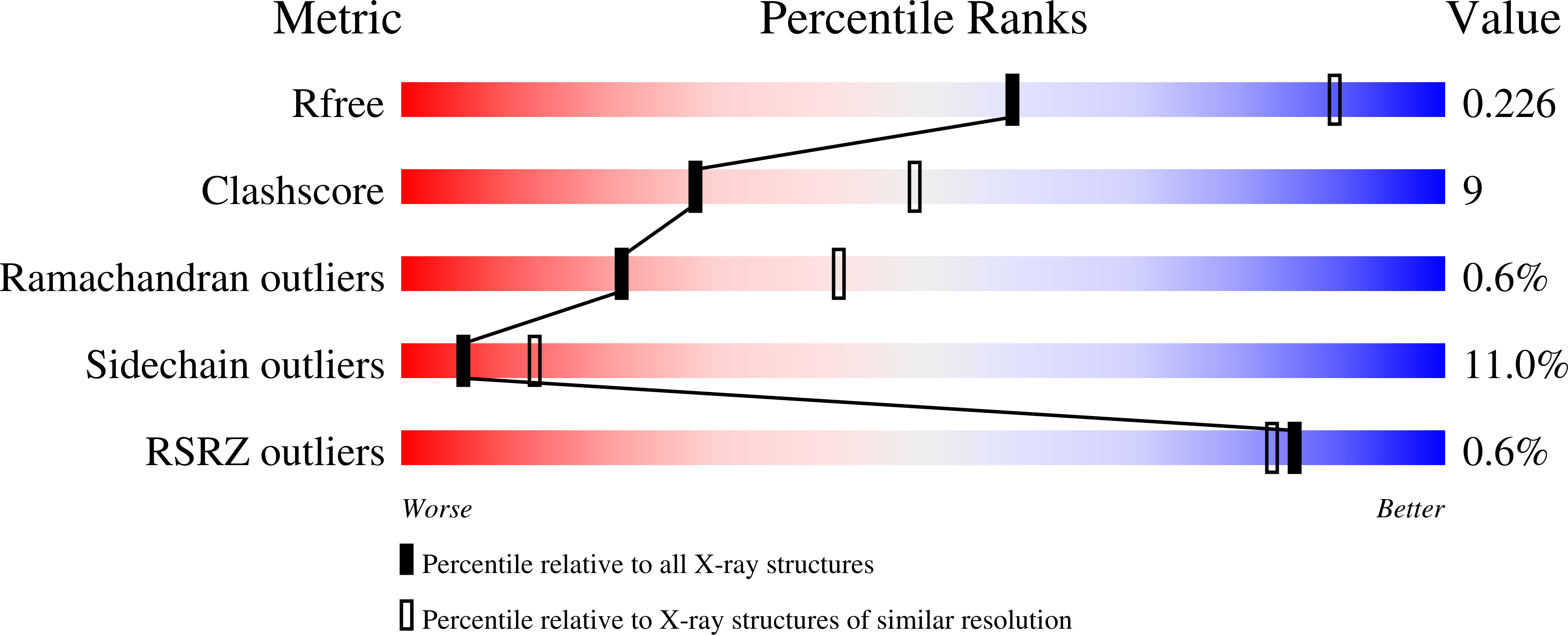
Experiments and Validation Details
wwPDB Validation report is not available for this entry.
X-ray source:
SPRING-8 BEAMLINE BL41XU
Spacegroup:
P212121
Expression system: Escherichia coli
