

-[alpha-D-mannopyranose-(1-6)]alpha-D-mannopyranose-(1-4)-2-acetamido-2-deoxy-beta-D-glucopyranose-(1-4)-[alpha-L-fucopyranose-(1-6)]2-acetamido-2-deoxy-beta-D-glucopyranose</span>;</li> <li class='image_legend_li'>8 copies of <span class='highlight'>PROTOPORPHYRIN IX CONTAINING FE</span>;</li> <li class='image_legend_li'>8 copies of <span class='highlight'>OXYGEN MOLECULE</span>;</li> <li class='image_legend_li'>2 copies of <span class='highlight'>CALCIUM ION</span>;</li> <li class='image_legend_li'>1 copy of <span class='highlight'>water</span>.</li></ul>")
-[alpha-D-mannopyranose-(1-6)]alpha-D-mannopyranose-(1-4)-2-acetamido-2-deoxy-beta-D-glucopyranose-(1-4)-[alpha-L-fucopyranose-(1-6)]2-acetamido-2-deoxy-beta-D-glucopyranose</span>;</li> <li class='image_legend_li'>8 copies of <span class='highlight'>PROTOPORPHYRIN IX CONTAINING FE</span>;</li> <li class='image_legend_li'>8 copies of <span class='highlight'>OXYGEN MOLECULE</span>;</li> <li class='image_legend_li'>2 copies of <span class='highlight'>CALCIUM ION</span>;</li> <li class='image_legend_li'>1 copy of <span class='highlight'>water</span>.</li></ul>")
-[alpha-D-mannopyranose-(1-6)]alpha-D-mannopyranose-(1-4)-2-acetamido-2-deoxy-beta-D-glucopyranose-(1-4)-[alpha-L-fucopyranose-(1-6)]2-acetamido-2-deoxy-beta-D-glucopyranose</span>;</li> <li class='image_legend_li'>8 copies of <span class='highlight'>PROTOPORPHYRIN IX CONTAINING FE</span>;</li> <li class='image_legend_li'>8 copies of <span class='highlight'>OXYGEN MOLECULE</span>;</li> <li class='image_legend_li'>2 copies of <span class='highlight'>CALCIUM ION</span>;</li> <li class='image_legend_li'>1 copy of <span class='highlight'>water</span>.</li></ul>")
Function and Biology Details
Biochemical function:
Biological process:
Cellular component:
Structure analysis Details
Assembly composition:
hetero 24-mer (preferred)
Assembly name:
Extracellular globin (preferred)
PDBe Complex ID:
PDB-CPX-197402 (preferred)
Entry contents:
4 distinct polypeptide molecules
Macromolecules (5 distinct):
Extracellular globin
Molecule details ›
Chains: A, E
Length: 146 amino acids
Theoretical weight: 16.37 KDa
Source organism: Lamellibrachia satsuma
UniProt:
Sequence domains: Globin
Structure domains: Globins
Length: 146 amino acids
Theoretical weight: 16.37 KDa
Source organism: Lamellibrachia satsuma
UniProt:
- Canonical:
 S0BBU7 (Residues: 20-165; Coverage: 100%)
Sequence domains: Globin
Structure domains: Globins
Extracellular globin
Extracellular globin
Extracellular globin
Ligands and Environments


No modified residues
Experiments and Validation Details
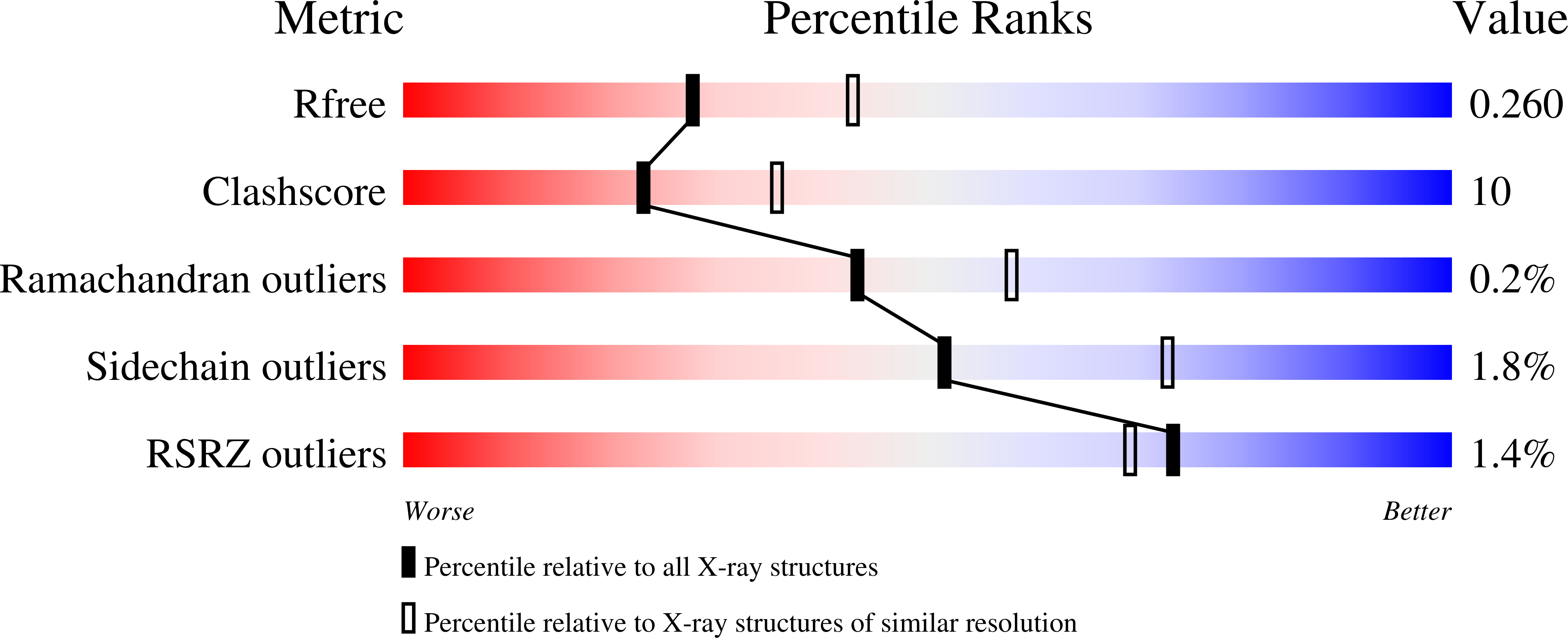
wwPDB Validation report is not available for this entry.
X-ray source:
SPRING-8 BEAMLINE BL44XU
Spacegroup:
P63
