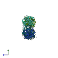
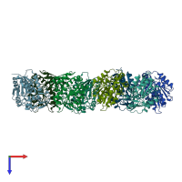
3x3h

X-ray diffraction
2.88Å resolution

Crystal Structure of the Manihot esculenta Hydroxynitrile Lyase (MeHNL) 3KP (K176P, K199P, K224P) triple mutant

Released:
Source organism: Manihot esculenta
Entry authors: Cielo CBC, Yamane T, Asano Y, Dadashipour M, Suzuki A, Mizushima T, Komeda H, Okazaki S

Function and Biology Details

Reaction catalysed:
An aliphatic (S)-hydroxynitrile = cyanide + an aliphatic aldehyde or ketone
Biochemical function:
Cellular component:
  • not assigned

Structure analysis Details

Assembly composition:
homo dimer (preferred)
PDBe Complex ID:
PDB-CPX-156605 (preferred)
Entry contents:
1 distinct polypeptide molecule
Macromolecule:
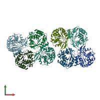
(S)-hydroxynitrile lyase Chains: A, B, C, D, E, F, G, H
Molecule details ›
Chains: A, B, C, D, E, F, G, H
Length: 258 amino acids
Theoretical weight: 29.31 KDa
Source organism: Manihot esculenta
Expression system: Escherichia coli
UniProt:
  • Canonical: P52705 (Residues: 1-258; Coverage: 100%)
Gene name: HNL
Sequence domains: alpha/beta hydrolase fold
Structure domains: alpha/beta hydrolase

Ligands and Environments

No bound ligands
No modified residues

Experiments and Validation Details

Entry percentile scores
X-ray source: RIGAKU FR-E DW
Spacegroup: P21
Unit cell:
a: 85.04Å b: 84.21Å c: 134.9Å
α: 90° β: 90.2° γ: 90°
R-values:
R R work R free
0.206 0.204 0.238
Expression system: Escherichia coli