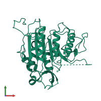
4afp

X-ray diffraction
2.1Å resolution

The structure of metacaspase 2 from T. brucei determined in the presence of Samarium

Released:
Model geometry
Fit model/data
Source organism: Trypanosoma brucei
Primary publication:
Crystal structure of a Trypanosoma brucei metacaspase.
Proc Natl Acad Sci U S A 109 7469-74 (2012)
PMID: 22529389

Function and Biology Details

Structure analysis Details

Assembly composition:
monomeric (preferred)
Assembly name:
PDBe Complex ID:
PDB-CPX-176656 (preferred)
Entry contents:
1 distinct polypeptide molecule
Macromolecule:
Metacaspase-2 Chain: A
Molecule details ›
Chain: A
Length: 367 amino acids
Theoretical weight: 39.94 KDa
Source organism: Trypanosoma brucei
Expression system: Escherichia coli BL21(DE3)
UniProt:
  • Canonical: Q585F3 (Residues: 1-347; Coverage: 100%)
Gene names: MCA2, Tb927.6.940
Sequence domains: Caspase domain

Ligands and Environments

1 bound ligand:
No modified residues

Experiments and Validation Details

wwPDB Validation report is not available for this entry.
X-ray source: DIAMOND BEAMLINE I04
Spacegroup: C2
Unit cell:
a: 78.217Å b: 49.787Å c: 85.224Å
α: 90° β: 117.1° γ: 90°
R-values:
R R work R free
0.184 0.181 0.238
Expression system: Escherichia coli BL21(DE3)