4atx

Electron Microscopy
8.2Å resolution

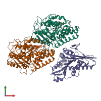
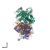
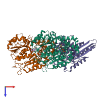
Rigor kinesin motor domain with an ordered neck-linker, docked on tubulin dimer, modelled into the 8A cryo-EM map of doublecortin- microtubules decorated with kinesin

Released:

Function and Biology Details

Structure analysis Details

Assembly composition:
hetero trimer (preferred)
PDBe Complex ID:
PDB-CPX-173620 (preferred)
Entry contents:
3 distinct polypeptide molecules
Macromolecules (3 distinct):
Tubulin beta-2B chain Chain: A
Molecule details ›
Chain: A
Length: 445 amino acids
Theoretical weight: 49.91 KDa
Source organism: Bos taurus
UniProt:
  • Canonical: Q6B856 (Residues: 1-445; Coverage: 100%)
Gene name: TUBB2B
Sequence domains:
Tubulin alpha-1D chain Chain: B
Molecule details ›
Chain: B
Length: 452 amino acids
Theoretical weight: 50.24 KDa
Source organism: Bos taurus
UniProt:
  • Canonical: Q2HJ86 (Residues: 1-452; Coverage: 100%)
Gene name: TUBA1D
Sequence domains:
Kinesin-1 heavy chain Chain: C
Molecule details ›
Chain: C
Length: 340 amino acids
Theoretical weight: 38.14 KDa
Source organism: Rattus norvegicus
Expression system: Escherichia coli
UniProt:
  • Canonical: Q2PQA9 (Residues: 1-340; Coverage: 35%)
Gene names: Khc, Kif5b
Sequence domains: Kinesin motor domain

Ligands and Environments

2 bound ligands:
No modified residues

Experiments and Validation Details

Entry percentile scores
Resolution: 8.2Å
Relevant EMDB volumes: EMD-2098
Expression system: Escherichia coli