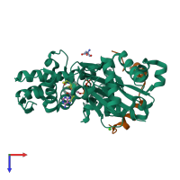
4b1u

X-ray diffraction
2Å resolution

Structure of the Phactr1 RPEL domain and RPEL motif directed assemblies with G-actin reveal the molecular basis for actin binding cooperativity.

Released:

Function and Biology Details

Structure analysis Details

Assembly composition:
hetero dimer (preferred)
PDBe Complex ID:
PDB-CPX-159298 (preferred)
Entry contents:
2 distinct polypeptide molecules
Macromolecules (2 distinct):
Actin, alpha skeletal muscle Chain: B
Molecule details ›
Chain: B
Length: 376 amino acids
Theoretical weight: 41.97 KDa
Source organism: Oryctolagus cuniculus
UniProt:
  • Canonical: P68134 (Residues: 2-377; Coverage: 100%)
Gene names: Acta, Acta1
Sequence domains: Actin
Structure domains:
Phosphatase and actin regulator 1 Chain: M
Molecule details ›
Chain: M
Length: 32 amino acids
Theoretical weight: 3.84 KDa
Source organism: Oryctolagus cuniculus
Expression system: Not provided
UniProt:
  • Canonical: Q2M3X8 (Residues: 133-164; Coverage: 6%)
Gene name: Phactr1
Sequence domains: RPEL repeat

Ligands and Environments

No modified residues

Experiments and Validation Details

Entry percentile scores
X-ray source: ESRF
Spacegroup: P3221
Unit cell:
a: 77.6Å b: 77.6Å c: 128.23Å
α: 90° β: 90° γ: 120°
R-values:
R R work R free
0.197 0.195 0.236
Expression system: Not provided