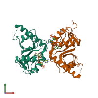
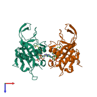
4b5p

X-ray diffraction
1.6Å resolution

Crystal structure of human alpha tubulin acetyltransferase catalytic domain Q58A variant

Released:
Source organism: Homo sapiens
Primary publication:
Atomic resolution structure of human α-tubulin acetyltransferase bound to acetyl-CoA.
Proc Natl Acad Sci U S A 109 19649-54 (2012)
PMID: 23071318

Function and Biology Details

Reaction catalysed:
Acetyl-CoA + [alpha-tubulin]-L-lysine = CoA + [alpha-tubulin]-N(6)-acetyl-L-lysine
Biological process:
Cellular component:

Structure analysis Details

Assembly composition:
hetero dimer (preferred)
PDBe Complex ID:
PDB-CPX-178274 (preferred)
Entry contents:
2 distinct polypeptide molecules
Macromolecules (2 distinct):
Alpha-tubulin N-acetyltransferase 1 Chain: A
Molecule details ›
Chain: A
Length: 200 amino acids
Theoretical weight: 22.79 KDa
Source organism: Homo sapiens
Expression system: Escherichia coli BL21(DE3)
UniProt:
  • Canonical: Q5SQI0 (Residues: 1-196; Coverage: 47%)
Gene names: ATAT1, C6orf134, MEC17, Nbla00487
Sequence domains: GNAT acetyltransferase, Mec-17
Structure domains: Aminopeptidase
Alpha-tubulin N-acetyltransferase 1 Chain: B
Molecule details ›
Chain: B
Length: 200 amino acids
Theoretical weight: 22.8 KDa
Source organism: Homo sapiens
Expression system: Escherichia coli BL21(DE3)
UniProt:
  • Canonical: Q5SQI0 (Residues: 1-196; Coverage: 47%)
Gene names: ATAT1, C6orf134, MEC17, Nbla00487
Sequence domains: GNAT acetyltransferase, Mec-17
Structure domains: Aminopeptidase

Ligands and Environments


Cofactor: Ligand ACO 2 x ACO
1 bound ligand:
No modified residues

Experiments and Validation Details

Entry percentile scores
X-ray source: SLS BEAMLINE X06DA
Spacegroup: P212121
Unit cell:
a: 36.76Å b: 110.87Å c: 113.58Å
α: 90° β: 90° γ: 90°
R-values:
R R work R free
0.186 0.185 0.222
Expression system: Escherichia coli BL21(DE3)