4dam

X-ray diffraction
1.7Å resolution

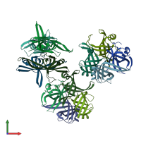
Crystal structure of small single-stranded DNA-binding protein from Streptomyces coelicolor

Released:

Function and Biology Details

Biochemical function:
Biological process:
Cellular component:
  • not assigned

Structure analysis Details

Assembly composition:
homo tetramer (preferred)
PDBe Complex ID:
PDB-CPX-192173 (preferred)
Entry contents:
1 distinct polypeptide molecule
Macromolecule:
Single-stranded DNA-binding protein 1 Chains: A, B, C, D, E, F, G, H, I, J, K, L
Molecule details ›
Chains: A, B, C, D, E, F, G, H, I, J, K, L
Length: 128 amino acids
Theoretical weight: 14.12 KDa
Source organism: Streptomyces coelicolor
Expression system: Escherichia coli
UniProt:
  • Canonical: Q9KYI9 (Residues: 1-116; Coverage: 74%)
Gene names: SCC61A.04c, SCO2683, ssb1
Sequence domains: Single-strand binding protein family
Structure domains: Nucleic acid-binding proteins

Ligands and Environments

No bound ligands
No modified residues

Experiments and Validation Details

Entry percentile scores
X-ray source: ESRF BEAMLINE BM14
Spacegroup: P32
Unit cell:
a: 150.004Å b: 150.004Å c: 54.7Å
α: 90° β: 90° γ: 120°
R-values:
R R work R free
0.155 0.153 0.192
Expression system: Escherichia coli