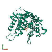
4dkb

X-ray diffraction
1.83Å resolution

Crystal Structure of Trypanosoma brucei dUTPase with dUpNp and Ca2+

Released:
Source organism: Trypanosoma brucei
Primary publication:
On the catalytic mechanism of dimeric dUTPases.
Biochem J 456 81-8 (2013)
PMID: 24001052

Function and Biology Details

Reaction catalysed:
dUTP + H(2)O = dUMP + diphosphate
Biochemical function:
Biological process:
  • not assigned
Cellular component:
Sequence domain:

Structure analysis Details

Assembly composition:
homo dimer (preferred)
PDBe Complex ID:
PDB-CPX-176544 (preferred)
Entry contents:
1 distinct polypeptide molecule
Macromolecule:
Deoxyuridine triphosphatase, putative Chain: A
Molecule details ›
Chain: A
Length: 290 amino acids
Theoretical weight: 32.2 KDa
Source organism: Trypanosoma brucei
Expression system: Escherichia coli BL21(DE3)
UniProt:
  • Canonical: Q57ZH3 (Residues: 1-287; Coverage: 100%)
Gene names: Tb07.27E10.390, Tb927.7.5160
Sequence domains: dUTPase
Structure domains: Type II deoxyuridine triphosphatase

Ligands and Environments

2 bound ligands:
No modified residues

Experiments and Validation Details

Entry percentile scores
X-ray source: ESRF BEAMLINE ID29
Spacegroup: P43212
Unit cell:
a: 68.47Å b: 68.47Å c: 123.87Å
α: 90° β: 90° γ: 90°
R-values:
R R work R free
0.17 0.169 0.198
Expression system: Escherichia coli BL21(DE3)