4eun

X-ray diffraction
1.6Å resolution

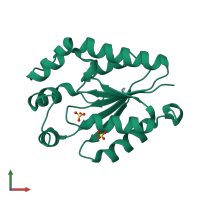
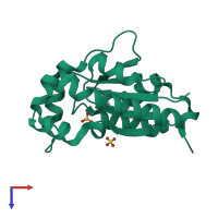
Crystal structure of a sugar kinase (Target EFI-502144 from Janibacter sp. HTCC2649), unliganded structure

Released:
Source organism: Janibacter sp. HTCC2649
Entry authors: Vetting MW, Toro R, Bhosle R, Al Obaidi NF, Morisco LL, Wasserman SR, Sojitra S, Washington E, Scott Glenn A, Chowdhury S, Evans B, Hammonds J, Hillerich B, Love J, Seidel RD, Imker HJ, Gerlt JA, Almo SC, Enzyme Function Initiative (EFI)

Function and Biology Details

Reaction catalysed:
ATP + D-gluconate = ADP + 6-phospho-D-gluconate
Biochemical function:
Biological process:
Cellular component:

Structure analysis Details

Assembly composition:
homo dimer (preferred)
Assembly name:
PDBe Complex ID:
PDB-CPX-107247 (preferred)
Entry contents:
1 distinct polypeptide molecule
Macromolecule:
Gluconokinase Chain: A
Molecule details ›
Chain: A
Length: 200 amino acids
Theoretical weight: 22.07 KDa
Source organism: Janibacter sp. HTCC2649
Expression system: Escherichia coli BL21(DE3)
UniProt:
  • Canonical: A3TPB6 (Residues: 1-177; Coverage: 100%)
Gene name: JNB_15458
Sequence domains: AAA domain
Structure domains: P-loop containing nucleotide triphosphate hydrolases

Ligands and Environments

1 bound ligand:
No modified residues

Experiments and Validation Details

Entry percentile scores
X-ray source: APS BEAMLINE 31-ID
Spacegroup: P3221
Unit cell:
a: 51.088Å b: 51.088Å c: 119.616Å
α: 90° β: 90° γ: 120°
R-values:
R R work R free
0.187 0.185 0.223
Expression system: Escherichia coli BL21(DE3)