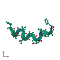
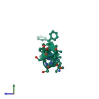
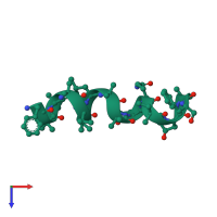
4g13

X-ray diffraction
0.8Å resolution

Crystal structure of samarosporin I at 100K

Released:
Source organism: samarospora rostrup
Primary publication:
The crystal structure of samarosporin I at atomic resolution.
J Pept Sci 18 678-84 (2012)
PMID: 23019149

Function and Biology Details

Biochemical function:
  • not assigned
Biological process:
  • not assigned
Cellular component:
  • not assigned

Structure analysis Details

Assembly composition:
monomeric (preferred)
Assembly name:
PDBe Complex ID:
PDB-CPX-164480 (preferred)
Entry contents:
1 distinct polypeptide molecule
Macromolecule:
SAMAROSPORIN I Chain: A
Molecule details ›
Chain: A
Length: 16 amino acids
Theoretical weight: 1.56 KDa
Source organism: samarospora rostrup

Ligands and Environments

No bound ligands
4 modified residues:

Experiments and Validation Details

Entry percentile scores
X-ray source: DIAMOND BEAMLINE I24
Spacegroup: C2
Unit cell:
a: 45.109Å b: 9.075Å c: 25.067Å
α: 90° β: 115.6° γ: 90°
R-values:
R R work R free
0.078 0.078 0.081