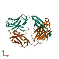
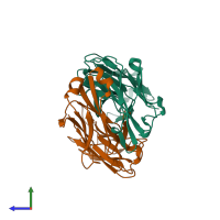
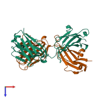
Function and Biology Details
Biochemical function:
- not assigned
Biological process:
- not assigned
Cellular component:
- not assigned
Structure analysis Details
Assembly composition:
hetero dimer (preferred)
Assembly name:
IG-heavy chain and IG-light kappa chain (preferred)
PDBe Complex ID:
PDB-CPX-213472 (preferred)
Entry contents:
2 distinct polypeptide molecules
Macromolecules (2 distinct):
Ligands and Environments
No bound ligands
No modified residues
Experiments and Validation Details
X-ray source:
SLS BEAMLINE X06SA
Spacegroup:
P212121
Expression system: Not provided


