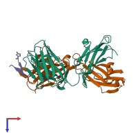
4gag

X-ray diffraction
1.8Å resolution

Structure of the broadly neutralizing antibody AP33 in complex with its HCV epitope (E2 residues 412-423)

Released:

Function and Biology Details

Structure analysis Details

Assembly composition:
hetero trimer (preferred)
PDBe Complex ID:
PDB-CPX-212302 (preferred)
Entry contents:
3 distinct polypeptide molecules
Macromolecules (3 distinct):
NEUTRALIZING ANTIBODY AP33 HEAVY CHAIN Chain: H
Molecule details ›
Chain: H
Length: 218 amino acids
Theoretical weight: 23.68 KDa
Source organism: Mus musculus
Structure domains: Immunoglobulins
NEUTRALIZING ANTIBODY AP33 LIGHT CHAIN Chain: L
Molecule details ›
Chain: L
Length: 218 amino acids
Theoretical weight: 23.92 KDa
Source organism: Mus musculus
Structure domains: Immunoglobulins
Envelope glycoprotein E2 Chain: P
Molecule details ›
Chain: P
Length: 12 amino acids
Theoretical weight: 1.37 KDa
Source organism: Hepatitis C virus (isolate Glasgow)
Expression system: Not provided
UniProt:
  • Canonical: Q5EG65 (Residues: 412-423; Coverage: 1%)

Ligands and Environments

1 bound ligand:
1 modified residue:

Experiments and Validation Details

Entry percentile scores
X-ray source: RIGAKU MICROMAX-007 HF
Spacegroup: C2
Unit cell:
a: 127.649Å b: 56.903Å c: 81.802Å
α: 90° β: 113.9° γ: 90°
R-values:
R R work R free
0.179 0.178 0.211
Expression system: Not provided