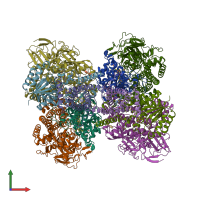
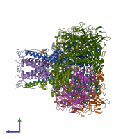
4gd3

X-ray diffraction
3.3Å resolution

Structure of E. coli hydrogenase-1 in complex with cytochrome b

Released:

Function and Biology Details

Structure analysis Details

Assemblies composition:
hetero pentamer
hetero dimer
hetero trimer (preferred)
PDBe Complex ID:
PDB-CPX-141993 (preferred)
Entry contents:
3 distinct polypeptide molecules
Macromolecules (3 distinct):
Hydrogenase-1 small chain Chains: Q, R, S, T
Molecule details ›
Chains: Q, R, S, T
Length: 335 amino acids
Theoretical weight: 36.82 KDa
Source organism: Escherichia coli K-12
Expression system: Escherichia coli
UniProt:
  • Canonical: P69739 (Residues: 46-372; Coverage: 100%)
Gene names: JW0954, b0972, hyaA
Sequence domains:
Structure domains:
Hydrogenase-1 large chain Chains: J, K, L, M
Molecule details ›
Chains: J, K, L, M
Length: 582 amino acids
Theoretical weight: 64.75 KDa
Source organism: Escherichia coli K-12
Expression system: Escherichia coli
UniProt:
  • Canonical: P0ACD8 (Residues: 1-582; Coverage: 98%)
Gene names: JW0955, b0973, hyaB
Sequence domains: Nickel-dependent hydrogenase
Structure domains: Cytochrome-c3 Hydrogenase, chain B
Probable Ni/Fe-hydrogenase 1 B-type cytochrome subunit Chains: A, B
Molecule details ›
Chains: A, B
Length: 235 amino acids
Theoretical weight: 27.63 KDa
Source organism: Escherichia coli K-12
Expression system: Escherichia coli
UniProt:
  • Canonical: P0AAM1 (Residues: 1-235; Coverage: 100%)
Gene names: JW0956, b0974, hyaC
Sequence domains: Prokaryotic cytochrome b561
Structure domains: Transmembrane di-heme cytochromes, Chain C

Ligands and Environments


Cofactor: Ligand HEM 2 x HEM
No modified residues

Experiments and Validation Details

Entry percentile scores
X-ray source: ESRF BEAMLINE ID29
Spacegroup: P212121
Unit cell:
a: 126Å b: 165.3Å c: 212.8Å
α: 90° β: 90° γ: 90°
R-values:
R R work R free
0.202 0.2 0.236
Expression system: Escherichia coli