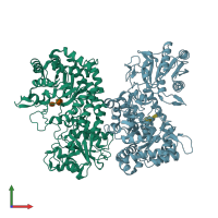
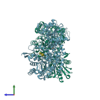
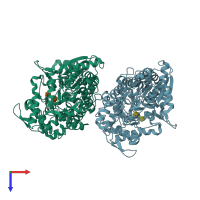
4gyk

X-ray diffraction
1.8Å resolution

Crystal structure of mutant (D318N) bacillus subtilis family 3 glycoside hydrolase (nagz) in complex with glcnac-murnac (space group P1211)

Released:

Function and Biology Details

Structure analysis Details

Assembly composition:
monomeric (preferred)
Assembly name:
PDBe Complex ID:
PDB-CPX-154110 (preferred)
Entry contents:
1 distinct polypeptide molecule
Macromolecules (2 distinct):
Beta-hexosaminidase Chains: A, B
Molecule details ›
Chains: A, B
Length: 648 amino acids
Theoretical weight: 71.23 KDa
Source organism: Bacillus subtilis subsp. subtilis str. 168
Expression system: Escherichia coli
UniProt:
  • Canonical: P40406 (Residues: 18-642; Coverage: 100%)
Gene names: BSU01660, nagZ, ybbD, yzbA
Sequence domains:
Structure domains:

Ligands and Environments

Carbohydrate polymer : NEW Components: MUB, NAG
No bound ligands
No modified residues

Experiments and Validation Details

Entry percentile scores
X-ray source: CLSI BEAMLINE 08ID-1
Spacegroup: P21
Unit cell:
a: 53.978Å b: 132.154Å c: 73.688Å
α: 90° β: 90.21° γ: 90°
R-values:
R R work R free
0.174 0.171 0.23
Expression system: Escherichia coli