


Function and Biology Details
Reaction catalysed:
dUMP + (6R)-5,10-methylene-5,6,7,8-tetrahydrofolate = 7,8-dihydrofolate +dTMP.
Biochemical function:
Biological process:
Cellular component:
Structure analysis Details
Assembly composition:
homo dimer (preferred)
Assembly name:
Thymidylate synthase (preferred)
PDBe Complex ID:
PDB-CPX-185655 (preferred)
Entry contents:
1 distinct polypeptide molecule
Macromolecule:
Thymidylate synthase
Molecule details ›
Chains: A, B
Length: 266 amino acids
Theoretical weight: 30.26 KDa
Source organism: Corynebacterium glutamicum ATCC 13032
Expression system: Escherichia coli
UniProt:
Sequence domains: Thymidylate synthase
Structure domains: Thymidylate synthase/dCMP hydroxymethylase domain
Length: 266 amino acids
Theoretical weight: 30.26 KDa
Source organism: Corynebacterium glutamicum ATCC 13032
Expression system: Escherichia coli
UniProt:
- Canonical:
 Q8NS38 (Residues: 1-266; Coverage: 100%)
Sequence domains: Thymidylate synthase
Structure domains: Thymidylate synthase/dCMP hydroxymethylase domain
Ligands and Environments
No bound ligands
No modified residues
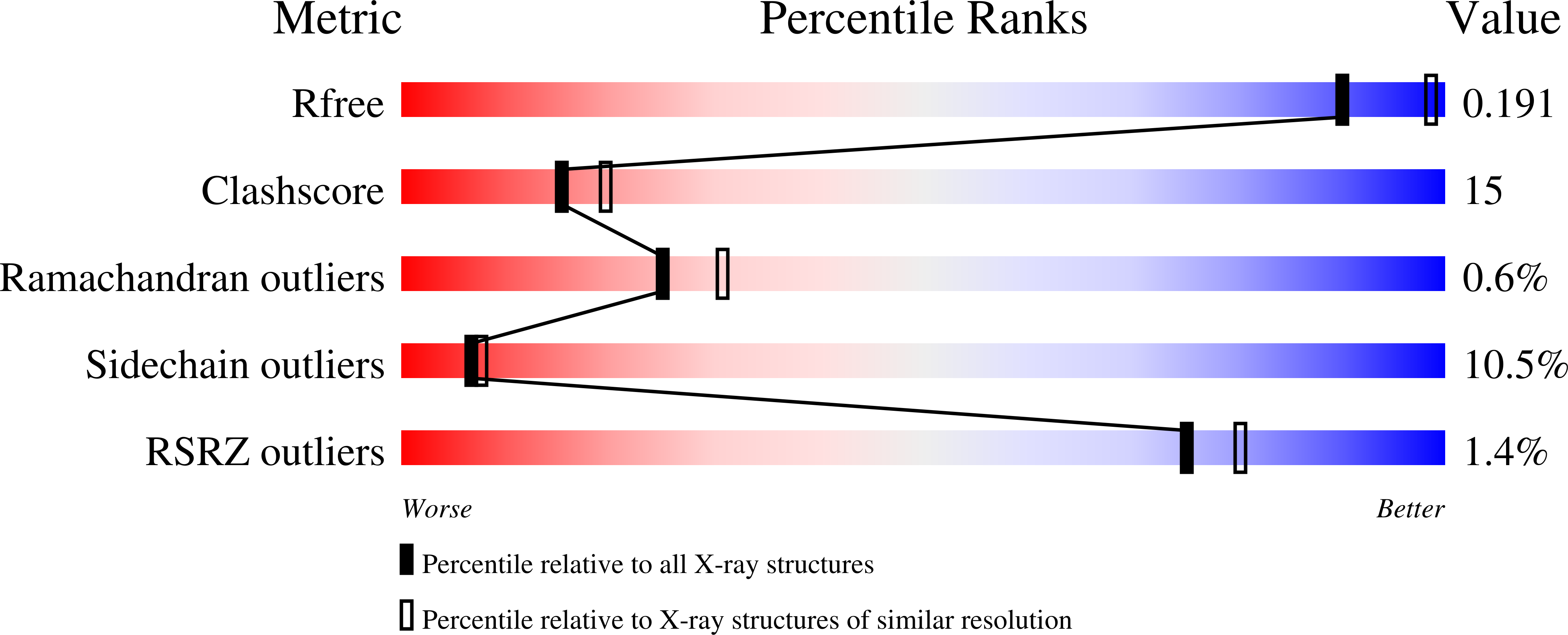
Experiments and Validation Details
wwPDB Validation report is not available for this entry.
X-ray source:
NSRRC BEAMLINE BL13B1
Spacegroup:
P21
Expression system: Escherichia coli
