


Function and Biology Details
Biochemical function:
Biological process:
Cellular component:
Structure analysis Details
Assembly composition:
homo heptamer (preferred)
Assembly name:
PDBe Complex ID:
PDB-CPX-108794 (preferred)
Entry contents:
1 distinct polypeptide molecule
Macromolecule:
Leukocidin/Hemolysin toxin domain-containing protein
Molecule details ›
Chains: A, B, C, D, E, F, G, H, I, J, K, L, M, N
Length: 296 amino acids
Theoretical weight: 33.51 KDa
Source organism: Clostridium perfringens
Expression system: Escherichia coli BL21(DE3)
UniProt:
Sequence domains: Leukocidin/Hemolysin toxin family
Structure domains: Leukocidin/porin MspA
Length: 296 amino acids
Theoretical weight: 33.51 KDa
Source organism: Clostridium perfringens
Expression system: Escherichia coli BL21(DE3)
UniProt:
- Canonical:
 A8ULG6 (Residues: 31-322; Coverage: 100%)
Sequence domains: Leukocidin/Hemolysin toxin family
Structure domains: Leukocidin/porin MspA
Ligands and Environments
No bound ligands
No modified residues
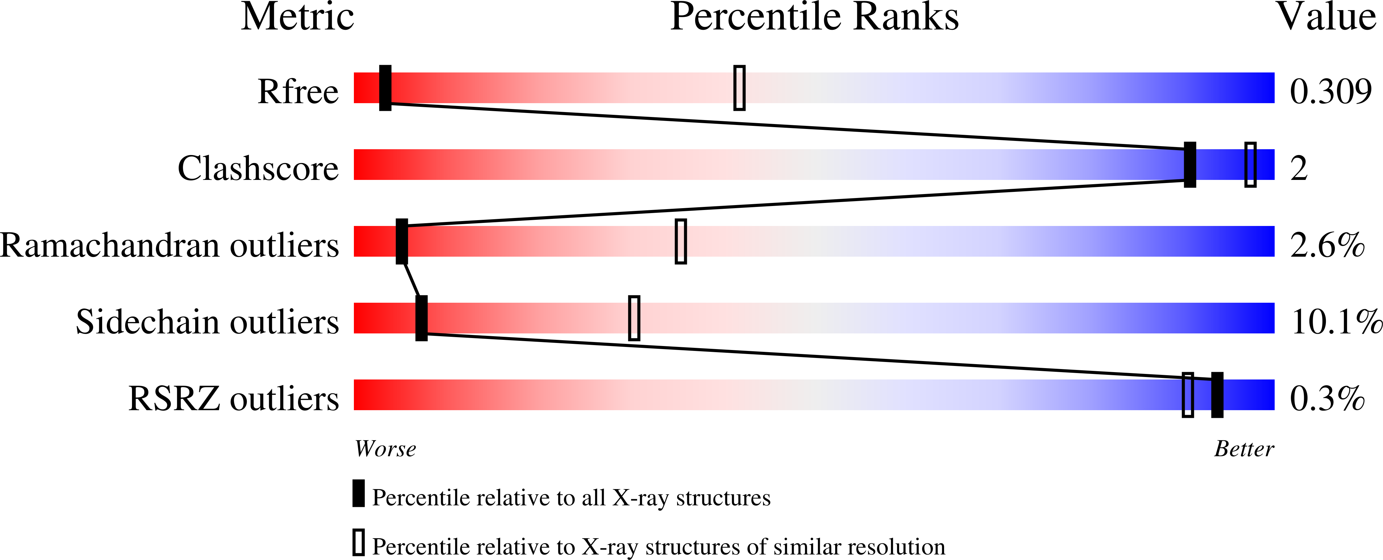
Experiments and Validation Details

X-ray source:
RIGAKU MICROMAX-007 HF
Spacegroup:
C2
Expression system: Escherichia coli BL21(DE3)