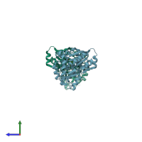
4hjx

X-ray diffraction
2.91Å resolution

Crystal structure of F2YRS complexed with F2Y

Released:
Primary publication:
A genetically encoded 19F NMR probe for tyrosine phosphorylation.
Angew Chem Int Ed Engl 52 3958-62 (2013)
PMID: 23450644

Function and Biology Details

Reaction catalysed:
ATP + L-tyrosine + tRNA(Tyr) = AMP + diphosphate + L-tyrosyl-tRNA(Tyr)
Biochemical function:
Biological process:
Cellular component:

Structure analysis Details

Assembly composition:
homo dimer (preferred)
Assembly name:
PDBe Complex ID:
PDB-CPX-176482 (preferred)
Entry contents:
1 distinct polypeptide molecule
Macromolecule:
Tyrosine--tRNA ligase Chains: A, B
Molecule details ›
Chains: A, B
Length: 314 amino acids
Theoretical weight: 36.05 KDa
Source organism: Methanocaldococcus jannaschii DSM 2661
Expression system: Escherichia coli
UniProt:
  • Canonical: Q57834 (Residues: 1-306; Coverage: 100%)
Gene names: MJ0389, tyrS
Sequence domains: tRNA synthetases class I (W and Y)
Structure domains:

Ligands and Environments

1 bound ligand:
No modified residues

Experiments and Validation Details

Entry percentile scores
X-ray source: RIGAKU MICROMAX-007 HF
Spacegroup: P43
Unit cell:
a: 101.307Å b: 101.307Å c: 71.779Å
α: 90° β: 90° γ: 90°
R-values:
R R work R free
0.214 0.211 0.268
Expression system: Escherichia coli