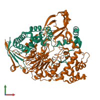
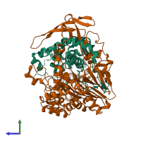
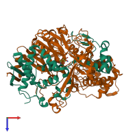
4hst

X-ray diffraction
1.57Å resolution

Crystal structure of a double mutant of a class III engineered cephalosporin acylase

Released:

Function and Biology Details

Structure analysis Details

Assembly composition:
hetero dimer (preferred)
PDBe Complex ID:
PDB-CPX-164554 (preferred)
Entry contents:
2 distinct polypeptide molecules
Macromolecules (2 distinct):
glutaryl-7-aminocephalosporanic acid acylase alpha chain Chain: A
Molecule details ›
Chain: A
Length: 229 amino acids
Theoretical weight: 24.85 KDa
Source organism: Pseudomonas
Expression system: Escherichia coli BL21(DE3)
Structure domains:
glutaryl-7-aminocephalosporanic acid acylase beta chain Chain: B
Molecule details ›
Chain: B
Length: 543 amino acids
Theoretical weight: 59.23 KDa
Source organism: Pseudomonas
Expression system: Escherichia coli BL21(DE3)
Structure domains:

Ligands and Environments

1 bound ligand:
No modified residues

Experiments and Validation Details

Entry percentile scores
X-ray source: AUSTRALIAN SYNCHROTRON BEAMLINE MX2
Spacegroup: P212121
Unit cell:
a: 68.385Å b: 77.845Å c: 191.99Å
α: 90° β: 90° γ: 90°
R-values:
R R work R free
0.121 0.119 0.162
Expression system: Escherichia coli BL21(DE3)