


Function and Biology Details
Reaction catalysed:
1-phosphatidyl-1D-myo-inositol = 1D-myo-inositol 1,2-cyclic phosphate + 1,2-diacyl-sn-glycerol
Biochemical function:
Biological process:
Cellular component:
- not assigned
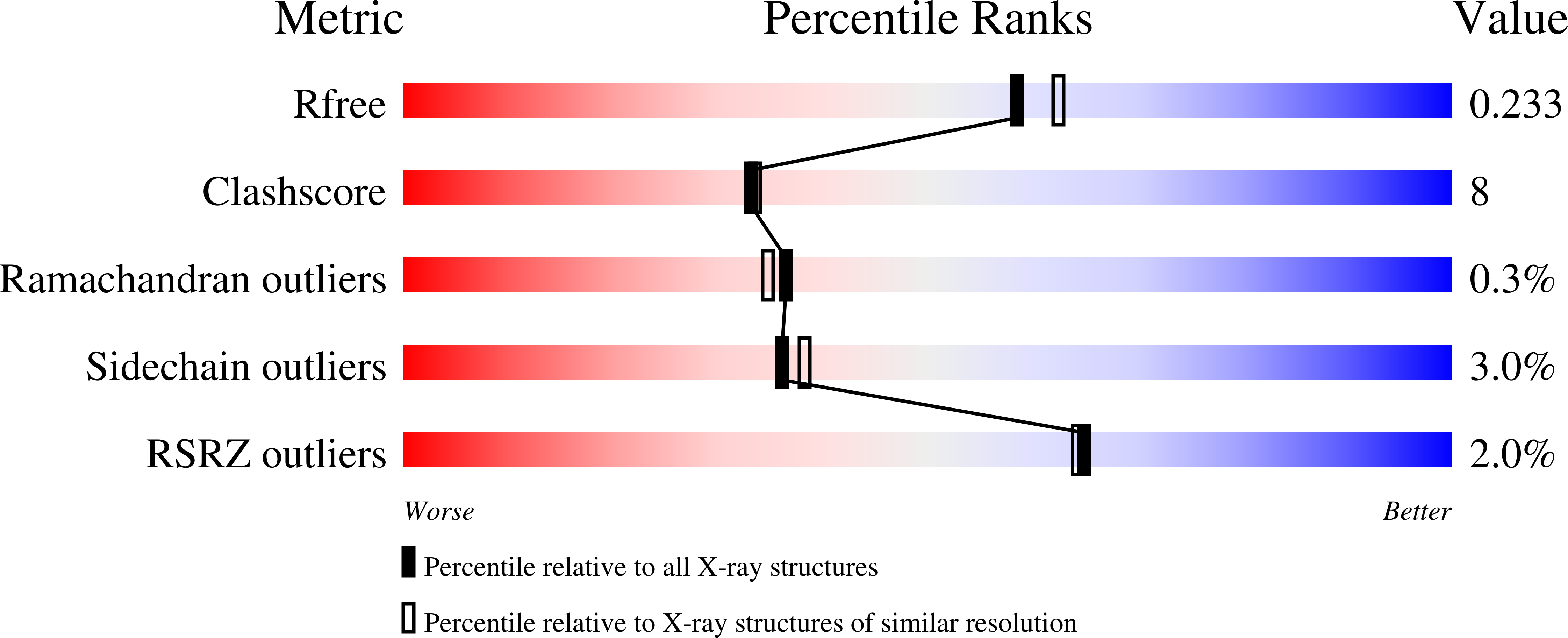
Structure analysis Details
Assembly composition:
monomeric (preferred)
Assembly name:
1-phosphatidylinositol phosphodiesterase (preferred)
PDBe Complex ID:
PDB-CPX-155335 (preferred)
Entry contents:
1 distinct polypeptide molecule
Macromolecule:
1-phosphatidylinositol phosphodiesterase
Molecule details ›
Chain: A
Length: 310 amino acids
Theoretical weight: 35.27 KDa
Source organism: Staphylococcus aureus subsp. aureus str. Newman
Expression system: Escherichia coli
UniProt:
Sequence domains: Phosphatidylinositol-specific phospholipase C, X domain
Structure domains: Phosphatidylinositol (PI) phosphodiesterase
Length: 310 amino acids
Theoretical weight: 35.27 KDa
Source organism: Staphylococcus aureus subsp. aureus str. Newman
Expression system: Escherichia coli
UniProt:
- Canonical:
 P45723 (Residues: 11-312; Coverage: 100%)
Sequence domains: Phosphatidylinositol-specific phospholipase C, X domain
Structure domains: Phosphatidylinositol (PI) phosphodiesterase


