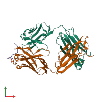
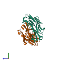
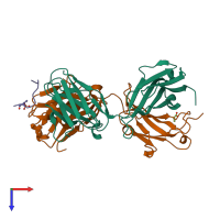
4jg1

X-ray diffraction
1.55Å resolution

Structure of phosphoserine/threonine (pSTAb) scaffold bound to pThr peptide

Released:
Source organism: Homo sapiens
Primary publication:
Nature-inspired design of motif-specific antibody scaffolds.
Nat Biotechnol 31 916-21 (2013)
PMID: 23955275

Function and Biology Details

Structure analysis Details

Assembly composition:
hetero trimer (preferred)
PDBe Complex ID:
PDB-CPX-211656 (preferred)
Entry contents:
3 distinct polypeptide molecules
Macromolecules (3 distinct):
Fab light chain Chain: L
Molecule details ›
Chain: L
Length: 214 amino acids
Theoretical weight: 23.3 KDa
Source organism: Homo sapiens
Expression system: Escherichia coli
Structure domains: Immunoglobulins
Fab heavy chain Chain: H
Molecule details ›
Chain: H
Length: 234 amino acids
Theoretical weight: 24.79 KDa
Source organism: Homo sapiens
Expression system: Escherichia coli
Structure domains: Immunoglobulins
Phosphopeptide Chain: P
Molecule details ›
Chain: P
Length: 12 amino acids
Theoretical weight: 1.42 KDa

Ligands and Environments

1 bound ligand:
1 modified residue:

Experiments and Validation Details

Entry percentile scores
X-ray source: ALS BEAMLINE 8.3.1
Spacegroup: P212121
Unit cell:
a: 43.807Å b: 95.591Å c: 119.817Å
α: 90° β: 90° γ: 90°
R-values:
R R work R free
0.152 0.151 0.174
Expression system: Escherichia coli