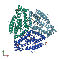
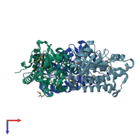
4k29

X-ray diffraction
1.66Å resolution

Crystal structure of an enoyl-CoA hydratase/isomerase from Xanthobacter autotrophicus Py2

Released:
Entry authors: Eswaramoorthy S, Chamala S, Evans B, Foti F, Gizzi A, Hillerich B, Kar A, Lafleur J, Seidel R, Villigas G, Zencheck W, Al Obaidi N, Almo SC, Swaminathan S, New York Structural Genomics Research Consortium (NYSGRC)

Function and Biology Details

Structure analysis Details

Assemblies composition:
homo hexamer
homo trimer (preferred)
PDBe Complex ID:
PDB-CPX-108258 (preferred)
Entry contents:
1 distinct polypeptide molecule
Macromolecule:
Enoyl-CoA hydratase/isomerase Chains: A, B, C
Molecule details ›
Chains: A, B, C
Length: 295 amino acids
Theoretical weight: 32.57 KDa
Source organism: Xanthobacter autotrophicus Py2
Expression system: Escherichia coli BL21
UniProt:
  • Canonical: A7IKN6 (Residues: 1-273; Coverage: 100%)
Gene name: Xaut_3350
Sequence domains: Enoyl-CoA hydratase/isomerase
Structure domains: 2-enoyl-CoA Hydratase; Chain A, domain 1

Ligands and Environments

2 bound ligands:
1 modified residue:

Experiments and Validation Details

Entry percentile scores
X-ray source: NSLS BEAMLINE X29A
Spacegroup: C2221
Unit cell:
a: 115.052Å b: 144.576Å c: 129.568Å
α: 90° β: 90° γ: 90°
R-values:
R R work R free
0.182 0.181 0.198
Expression system: Escherichia coli BL21