

-2-METHYL-2,4-PENTANEDIOL</span>;</li> <li class='image_legend_li'>1 copy of <span class='highlight'>D(-)-TARTARIC ACID</span>;</li> <li class='image_legend_li'>2 copies of <span class='highlight'>N-acetyl-alpha-muramic acid</span>;</li> <li class='image_legend_li'>1 copy of <span class='highlight'>water</span>.</li></ul>")
-2-METHYL-2,4-PENTANEDIOL</span>;</li> <li class='image_legend_li'>1 copy of <span class='highlight'>D(-)-TARTARIC ACID</span>;</li> <li class='image_legend_li'>2 copies of <span class='highlight'>N-acetyl-alpha-muramic acid</span>;</li> <li class='image_legend_li'>1 copy of <span class='highlight'>water</span>.</li></ul>")
-2-METHYL-2,4-PENTANEDIOL</span>;</li> <li class='image_legend_li'>1 copy of <span class='highlight'>D(-)-TARTARIC ACID</span>;</li> <li class='image_legend_li'>2 copies of <span class='highlight'>N-acetyl-alpha-muramic acid</span>;</li> <li class='image_legend_li'>1 copy of <span class='highlight'>water</span>.</li></ul>")
Function and Biology Details
Reactions catalysed:
an N(4)-(oligosaccharide-(1->3)-[oligosaccharide-(1->6)]-beta-D-Man-(1->4)-beta-D-GlcNAc-(1->4)-alpha-D-GlcNAc)-L-asparaginyl-[protein] +H2O = an oligosaccharide-(1->3)-[oligosaccharide-(1->6)]-beta-D-Man-(1->4)-D-GlcNAc + N(4)-(N-acetyl-beta-D-glucosaminyl)-L-asparaginyl-[protein].
Hydrolyzes the link between N-acetylmuramoyl residues and L-amino acidresidues in certain cell-wall glycopeptides.
Biochemical function:
Biological process:
Cellular component:
- not assigned
Structure analysis Details
Assemblies composition:
Assembly name:
Bifunctional autolysin and peptide (preferred)
PDBe Complex ID:
PDB-CPX-164695 (preferred)
Entry contents:
2 distinct polypeptide molecules
Macromolecules (2 distinct):
Bifunctional autolysin
Molecule details ›
Chains: A, B, C, D
Length: 225 amino acids
Theoretical weight: 25.27 KDa
Source organism: Staphylococcus aureus subsp. aureus NCTC 8325
Expression system: Escherichia coli BL21(DE3)
UniProt:
Sequence domains: N-acetylmuramoyl-L-alanine amidase
Structure domains: Peptidoglycan recognition protein-like
Length: 225 amino acids
Theoretical weight: 25.27 KDa
Source organism: Staphylococcus aureus subsp. aureus NCTC 8325
Expression system: Escherichia coli BL21(DE3)
UniProt:
- Canonical:
 Q2FZK7 (Residues: 198-421; Coverage: 18%)
Sequence domains: N-acetylmuramoyl-L-alanine amidase
Structure domains: Peptidoglycan recognition protein-like
Ligands and Environments


Experiments and Validation Details
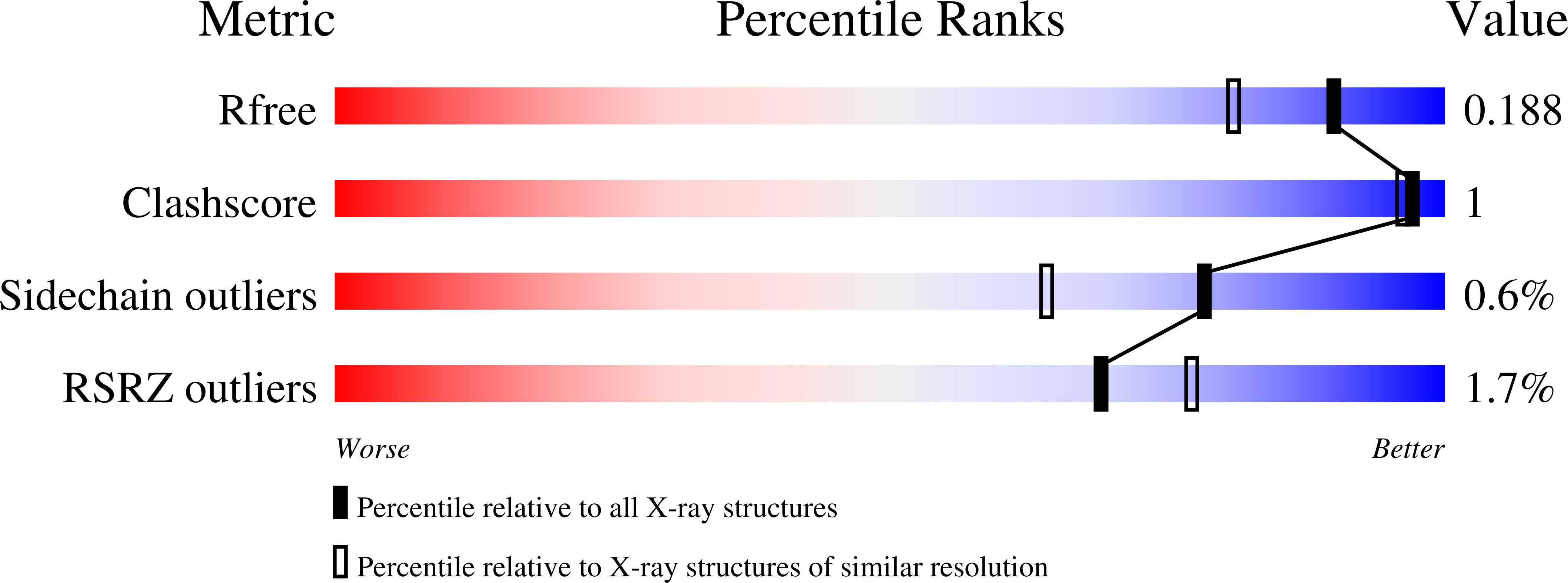
wwPDB Validation report is not available for this entry.
X-ray source:
SLS BEAMLINE X06SA
Spacegroup:
P21
Expression systems:
- Escherichia coli BL21(DE3)
- Not provided
