


Function and Biology Details
Reaction catalysed:
Hydrolyzes single-stranded DNA or mismatched double-stranded DNA andpolynucleotides, releasing free uracil.
Biochemical function:
Biological process:
Cellular component:
- not assigned
Structure analysis Details
Assemblies composition:
Assembly name:
PDBe Complex ID:
PDB-CPX-147057 (preferred)
Entry contents:
2 distinct polypeptide molecules
Macromolecules (2 distinct):
Uracil-DNA glycosylase
Molecule details ›
Chains: A, C, E, G, I, K, M, O
Length: 223 amino acids
Theoretical weight: 25.29 KDa
Source organism: Gadus morhua
Expression system: Escherichia coli
UniProt:
Sequence domains: Uracil DNA glycosylase superfamily
Structure domains: Uracil-DNA glycosylase-like domain
Length: 223 amino acids
Theoretical weight: 25.29 KDa
Source organism: Gadus morhua
Expression system: Escherichia coli
UniProt:
- Canonical:
 Q9I983 (Residues: 82-301; Coverage: 73%)
Sequence domains: Uracil DNA glycosylase superfamily
Structure domains: Uracil-DNA glycosylase-like domain
Uracil-DNA glycosylase inhibitor
Molecule details ›
Chains: B, D, F, H, J, L, N, P
Length: 84 amino acids
Theoretical weight: 9.48 KDa
Source organism: Bacillus phage PBS2
Expression system: Escherichia coli
UniProt:
Sequence domains: Uracil-DNA glycosylase inhibitor
Structure domains: Bacteriophage PBS2, uracil-glycosylase inhibitor
Length: 84 amino acids
Theoretical weight: 9.48 KDa
Source organism: Bacillus phage PBS2
Expression system: Escherichia coli
UniProt:
- Canonical: P14739 (Residues: 1-84; Coverage: 100%)
Sequence domains: Uracil-DNA glycosylase inhibitor
Structure domains: Bacteriophage PBS2, uracil-glycosylase inhibitor
Ligands and Environments
No bound ligands
No modified residues
Experiments and Validation Details
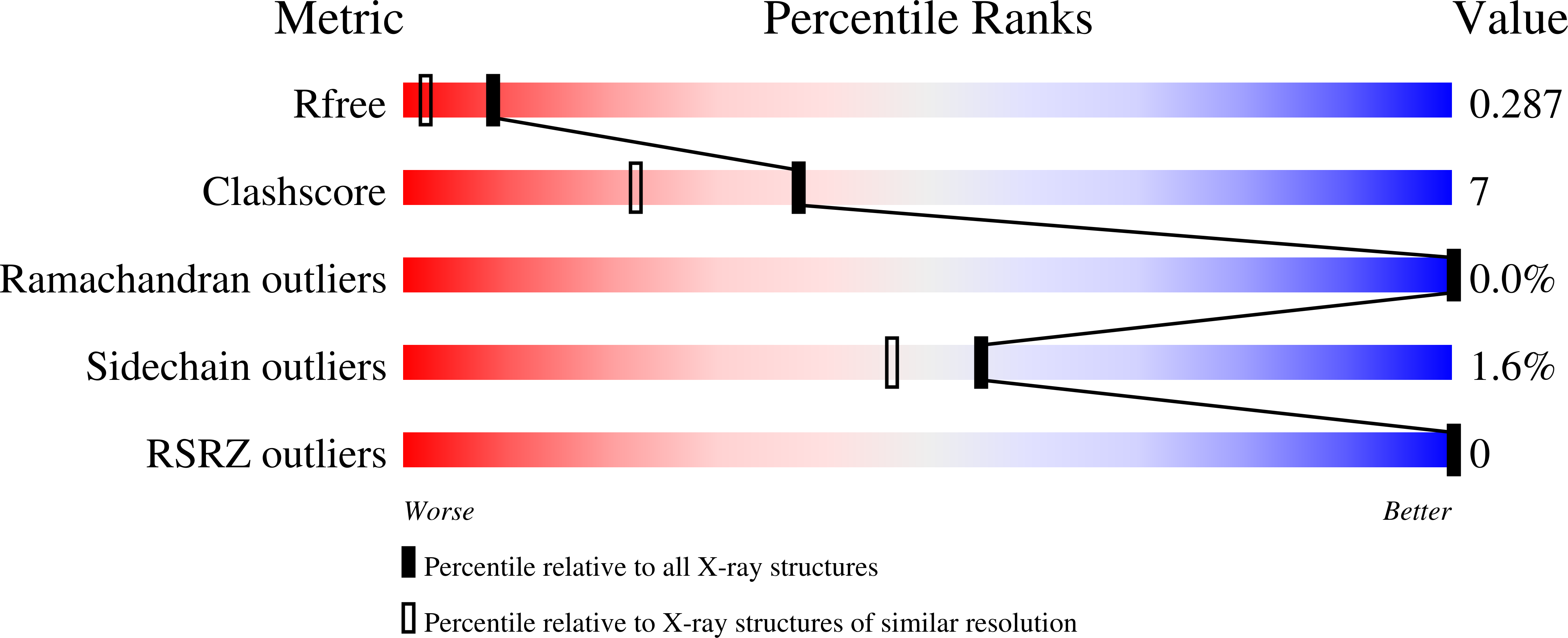
wwPDB Validation report is not available for this entry.
X-ray source:
ESRF BEAMLINE ID29
Spacegroup:
P21
Expression system: Escherichia coli
