


Function and Biology Details
Biochemical function:
- not assigned
Biological process:
Cellular component:
Structure analysis Details
Assembly composition:
monomeric (preferred)
Assembly name:
Beta-2-microglobulin (preferred)
PDBe Complex ID:
PDB-CPX-170033 (preferred)
Entry contents:
1 distinct polypeptide molecule
Macromolecule:
Beta-2-microglobulin
Molecule details ›
Chain: A
Length: 107 amino acids
Theoretical weight: 12.44 KDa
Source organism: Danio rerio
Expression system: Escherichia coli BL21(DE3)
UniProt:
Sequence domains: Immunoglobulin C1-set domain
Structure domains: Immunoglobulins
Length: 107 amino acids
Theoretical weight: 12.44 KDa
Source organism: Danio rerio
Expression system: Escherichia coli BL21(DE3)
UniProt:
- Canonical:
 Q04475 (Residues: 19-116; Coverage: 100%)
Sequence domains: Immunoglobulin C1-set domain
Structure domains: Immunoglobulins
Ligands and Environments
No bound ligands
No modified residues
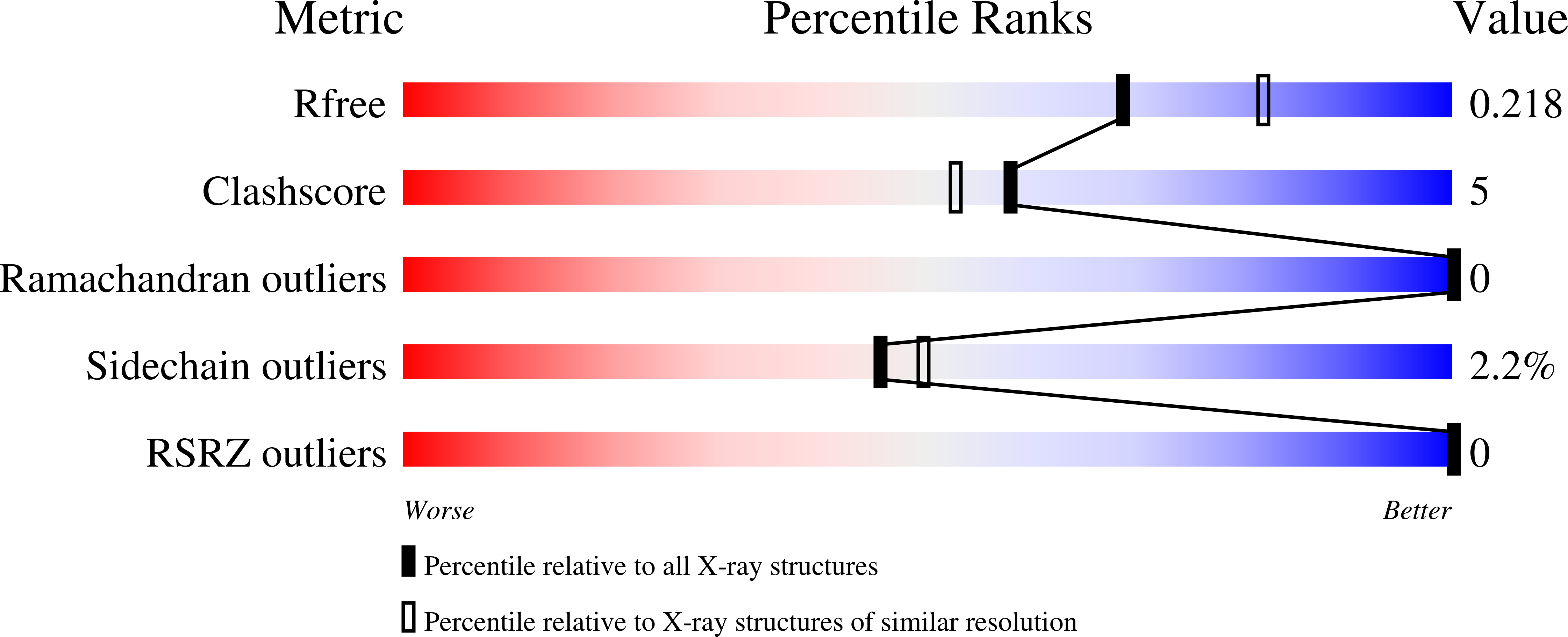
Experiments and Validation Details
wwPDB Validation report is not available for this entry.
X-ray source:
NSRRC BEAMLINE BL13B1
Spacegroup:
P212121
Expression system: Escherichia coli BL21(DE3)
