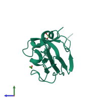
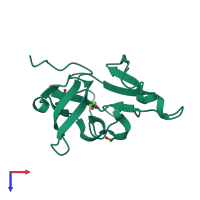
4me8

X-ray diffraction
2.27Å resolution

Crystal structure of a signal peptidase I (EF3073) from Enterococcus faecalis V583 at 2.27 A resolution

Released:
Source organism: Enterococcus faecalis V583
Entry author: Joint Center for Structural Genomics (JCSG)

Function and Biology Details

Reaction catalysed:
Cleavage of hydrophobic, N-terminal signal or leader sequences from secreted and periplasmic proteins.
Biochemical function:
Biological process:
Cellular component:

Structure analysis Details

Assembly composition:
monomeric (preferred)
Assembly name:
PDBe Complex ID:
PDB-CPX-182738 (preferred)
Entry contents:
1 distinct polypeptide molecule
Macromolecule:
Signal peptidase I Chain: A
Molecule details ›
Chain: A
Length: 151 amino acids
Theoretical weight: 17.16 KDa
Source organism: Enterococcus faecalis V583
Expression system: Escherichia coli
UniProt:
  • Canonical: Q82ZI3 (Residues: 29-178; Coverage: 84%)
Gene name: EF_3073
Sequence domains: Signal peptidase, peptidase S26
Structure domains: Umud Fragment, subunit A

Ligands and Environments

1 bound ligand:
1 modified residue:

Experiments and Validation Details

Entry percentile scores
X-ray source: SSRL BEAMLINE BL11-1
Spacegroup: P41212
Unit cell:
a: 37.488Å b: 37.488Å c: 169.046Å
α: 90° β: 90° γ: 90°
R-values:
R R work R free
0.216 0.212 0.258
Expression system: Escherichia coli