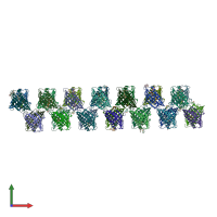
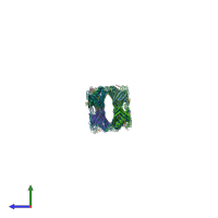
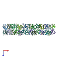
4n3e

X-ray diffraction
2.43Å resolution

Crystal structure of Hyp-1, a St John's wort PR-10 protein, in complex with 8-anilino-1-naphthalene sulfonate (ANS)

Released:

Function and Biology Details

Structure analysis Details

Assembly composition:
monomeric (preferred)
PDBe Complex ID:
PDB-CPX-184606 (preferred)
Entry contents:
1 distinct polypeptide molecule
Macromolecule:
Bet v I/Major latex protein domain-containing protein Chains: A, B, C, D, E, F, G, H, I, J, K, L, M, N, O, P, Q, R, S, T, U, V, W, X, Y, Z, a, b
Molecule details ›
Chains: A, B, C, D, E, F, G, H, I, J, K, L, M, N, O, P, Q, R, S, T, U, V, W, X, Y, Z, a, b
Length: 165 amino acids
Theoretical weight: 18.5 KDa
Source organism: Hypericum perforatum
Expression system: Escherichia coli BL21(DE3)
UniProt:
  • Canonical: Q8H1L1 (Residues: 1-159; Coverage: 100%)
Gene name: HYP1.1
Sequence domains: Pathogenesis-related protein Bet v 1 family
Structure domains: Alpha-D-Glucose-1,6-Bisphosphate; Chain A, domain 4

Ligands and Environments

3 bound ligands:
No modified residues

Experiments and Validation Details

Entry percentile scores
X-ray source: APS BEAMLINE 22-BM
Spacegroup: C2
Unit cell:
a: 146.29Å b: 146.29Å c: 298.56Å
α: 90° β: 90° γ: 90°
R-values:
R R work R free
0.224 0.223 0.278
Expression system: Escherichia coli BL21(DE3)