


Function and Biology Details
Reaction catalysed:
Endohydrolysis of (1->4)-beta-D-glucosidic linkages in cellulose, lichenin and cereal beta-D-glucans
Biochemical function:
Biological process:
Cellular component:
- not assigned
Structure analysis Details
Assembly composition:
monomeric (preferred)
Assembly name:
Endoglucanase (preferred)
PDBe Complex ID:
PDB-CPX-122685 (preferred)
Entry contents:
1 distinct polypeptide molecule
Macromolecule:
Endoglucanase
Molecule details ›
Chain: A
Length: 363 amino acids
Theoretical weight: 41.53 KDa
Source organism: Butyrivibrio proteoclasticus B316
Expression system: Escherichia coli
UniProt:
Sequence domains: Cellulase (glycosyl hydrolase family 5)
Structure domains: Glycosidases
Length: 363 amino acids
Theoretical weight: 41.53 KDa
Source organism: Butyrivibrio proteoclasticus B316
Expression system: Escherichia coli
UniProt:
- Canonical:
 E0RXM0 (Residues: 34-394; Coverage: 69%)
Sequence domains: Cellulase (glycosyl hydrolase family 5)
Structure domains: Glycosidases
Ligands and Environments
No bound ligands
No modified residues
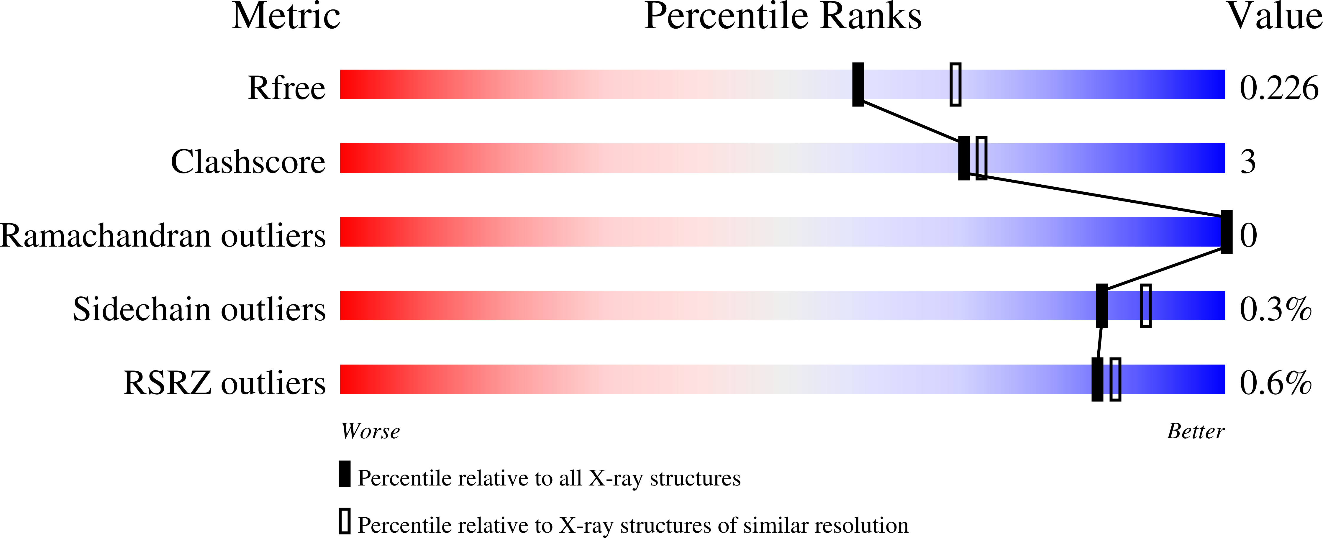
Experiments and Validation Details
wwPDB Validation report is not available for this entry.
X-ray source:
AUSTRALIAN SYNCHROTRON BEAMLINE MX1
Spacegroup:
P43212
Expression system: Escherichia coli
