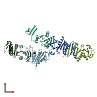
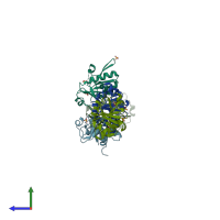
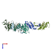
4nkr

X-ray diffraction
2.41Å resolution

The Crystal structure of Bacillus subtilis MobB

Released:
Entry authors: Choe J, Kim D, Choi S, Kim H

Function and Biology Details

Structure analysis Details

Assembly composition:
homo dimer (preferred)
PDBe Complex ID:
PDB-CPX-122701 (preferred)
Entry contents:
1 distinct polypeptide molecule
Macromolecule:
Molybdopterin-guanine dinucleotide biosynthesis protein B (MobB) domain-containing protein Chains: A, B, C, D, E
Molecule details ›
Chains: A, B, C, D, E
Length: 166 amino acids
Theoretical weight: 18.48 KDa
Source organism: Bacillus subtilis subsp. spizizenii str. W23
UniProt:
  • Canonical: E0U3U4 (Residues: 8-172; Coverage: 95%)
Gene names: BSUW23_07340, mobB
Sequence domains: Molybdopterin guanine dinucleotide synthesis protein B
Structure domains: P-loop containing nucleotide triphosphate hydrolases

Ligands and Environments

1 bound ligand:
No modified residues

Experiments and Validation Details

Entry percentile scores
X-ray source: PAL/PLS BEAMLINE 5C (4A)
Spacegroup: C2
Unit cell:
a: 225.528Å b: 42.107Å c: 93.62Å
α: 90° β: 100.99° γ: 90°
R-values:
R R work R free
0.231 0.227 0.321