


Function and Biology Details
Reaction catalysed:
a pyrimidine ribonucleoside 5'-phosphate + H2O = a pyrimidine nucleobase+ D-ribose 5-phosphate.
Biochemical function:
Biological process:
- not assigned
Cellular component:
- not assigned
Structure analysis Details
Assembly composition:
monomeric (preferred)
Assembly name:
AB hydrolase-1 domain-containing protein (preferred)
PDBe Complex ID:
PDB-CPX-124658 (preferred)
Entry contents:
1 distinct polypeptide molecule
Macromolecule:
AB hydrolase-1 domain-containing protein
Molecule details ›
Chains: A, B
Length: 320 amino acids
Theoretical weight: 37.09 KDa
Source organism: Priestia megaterium
Expression system: Escherichia coli BL21
UniProt:
Structure domains: alpha/beta hydrolase
Length: 320 amino acids
Theoretical weight: 37.09 KDa
Source organism: Priestia megaterium
Expression system: Escherichia coli BL21
UniProt:
- Canonical:
 G9BEX6 (Residues: 1-287; Coverage: 100%)
Structure domains: alpha/beta hydrolase
Ligands and Environments
No bound ligands
No modified residues
Experiments and Validation Details
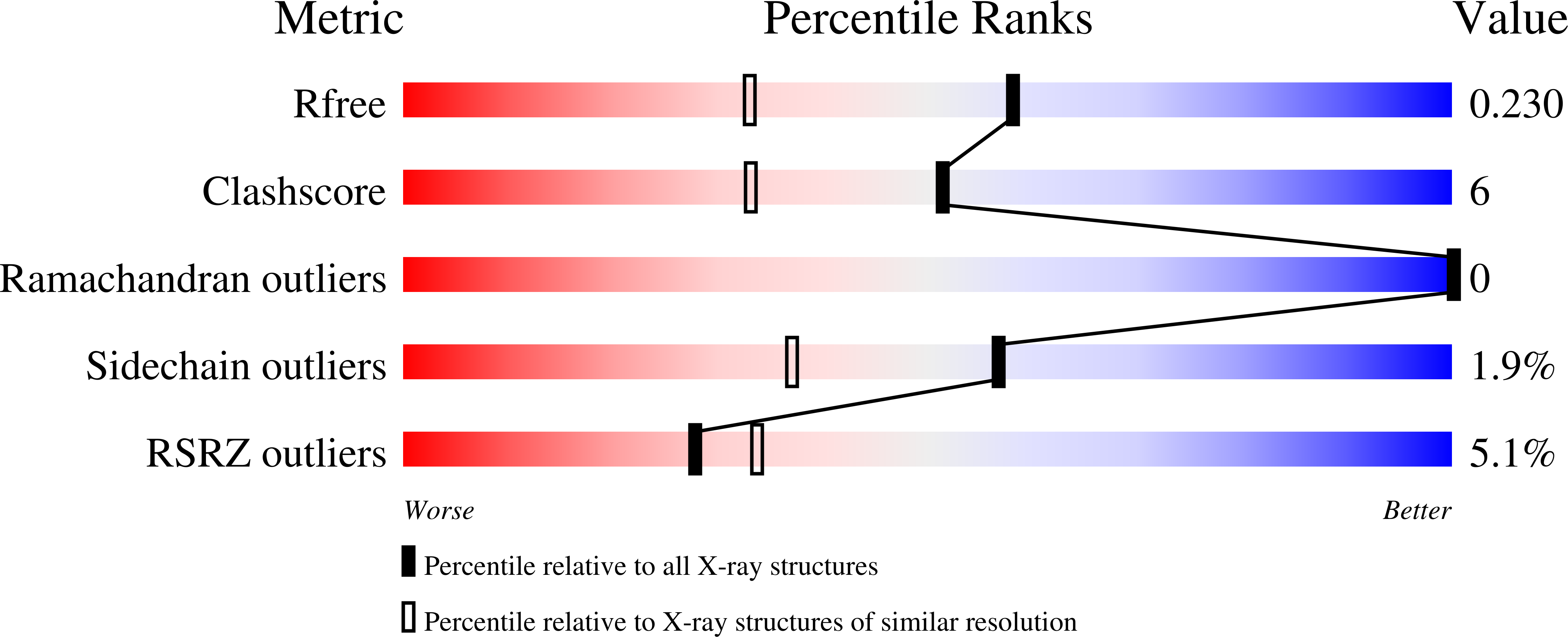
wwPDB Validation report is not available for this entry.
X-ray source:
SSRF BEAMLINE BL17U
Spacegroup:
P212121
Expression system: Escherichia coli BL21
