


Function and Biology Details
Biochemical function:
- not assigned
Biological process:
- not assigned
Cellular component:
- not assigned
Sequence domains:
Structure analysis Details
Assembly composition:
monomeric (preferred)
Assembly name:
AB hydrolase-1 domain-containing protein (preferred)
PDBe Complex ID:
PDB-CPX-179355 (preferred)
Entry contents:
1 distinct polypeptide molecule
Macromolecule:
AB hydrolase-1 domain-containing protein
Molecule details ›
Chains: A, B
Length: 298 amino acids
Theoretical weight: 34.56 KDa
Source organism: Rickettsia typhi str. Wilmington
Expression system: Escherichia coli
UniProt:
Sequence domains: alpha/beta hydrolase fold
Structure domains: alpha/beta hydrolase
Length: 298 amino acids
Theoretical weight: 34.56 KDa
Source organism: Rickettsia typhi str. Wilmington
Expression system: Escherichia coli
UniProt:
- Canonical:
 Q68WT4 (Residues: 1-290; Coverage: 100%)
Sequence domains: alpha/beta hydrolase fold
Structure domains: alpha/beta hydrolase
Ligands and Environments
No bound ligands
No modified residues
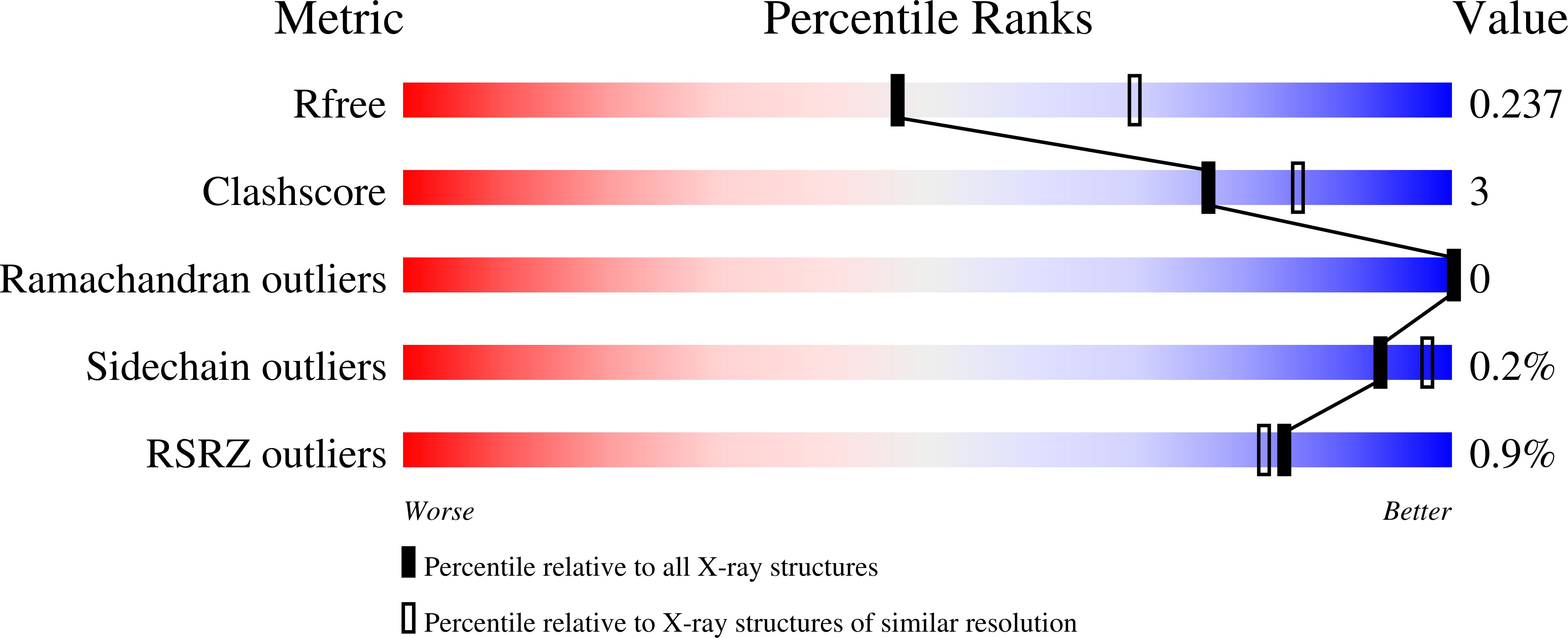
Experiments and Validation Details

X-ray source:
APS BEAMLINE 21-ID-F
Spacegroup:
C2
Expression system: Escherichia coli