

-3')</span>.</li></ul>")
-3')</span>.</li></ul>")
-3')</span>.</li></ul>")

Function and Biology Details
Biochemical function:
- not assigned
Biological process:
- not assigned
Cellular component:
- not assigned
Sequence domain:
Structure analysis Details
Assembly composition:
hetero hexamer (preferred)
Assembly name:
Replication restart protein DnaT and DNA (preferred)
PDBe Complex ID:
PDB-CPX-117951 (preferred)
Entry contents:
1 distinct polypeptide molecule
1 distinct DNA molecule
1 distinct DNA molecule
Macromolecules (2 distinct):
Replication restart protein DnaT
Molecule details ›
Chains: A, B, C, D, E
Length: 71 amino acids
Theoretical weight: 8.17 KDa
Source organism: Escherichia coli K-12
Expression system: Escherichia coli
UniProt:
Sequence domains: DnaT DNA-binding domain
Structure domains: Helicase, Ruva Protein; domain 3
Length: 71 amino acids
Theoretical weight: 8.17 KDa
Source organism: Escherichia coli K-12
Expression system: Escherichia coli
UniProt:
- Canonical:
 P0A8J2 (Residues: 84-154; Coverage: 40%)
Sequence domains: DnaT DNA-binding domain
Structure domains: Helicase, Ruva Protein; domain 3
Ligands and Environments
No bound ligands
No modified residues
Experiments and Validation Details
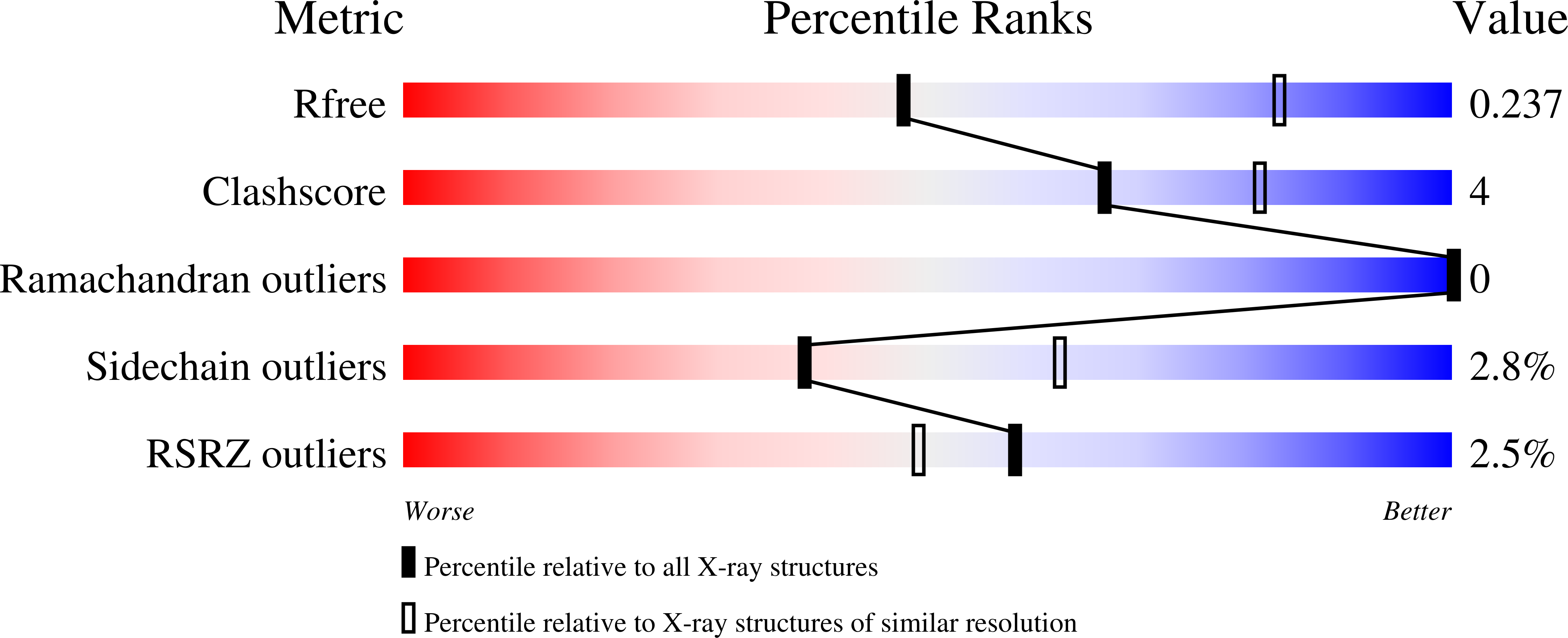
wwPDB Validation report is not available for this entry.
X-ray source:
SSRF BEAMLINE BL17U
Spacegroup:
P1
Expression systems:
- Escherichia coli
- Not provided
