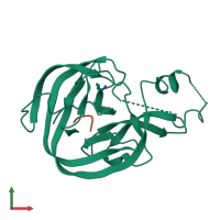
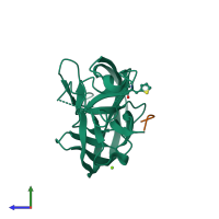
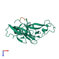
4oz6

X-ray diffraction
2.79Å resolution

Structure of the Branched Intermediate in Protein Splicing

Released:
Source organism: Mycobacterium xenopi
Primary publication:
Structure of the branched intermediate in protein splicing.
Proc Natl Acad Sci U S A 111 8422-7 (2014)
PMID: 24778214

Function and Biology Details

Reaction catalysed:
ATP-dependent breakage, passage and rejoining of double-stranded DNA
Biochemical function:
  • not assigned
Cellular component:
  • not assigned

Structure analysis Details

Assembly composition:
hetero dimer (preferred)
Assembly name:
PDBe Complex ID:
PDB-CPX-159830 (preferred)
Entry contents:
2 distinct polypeptide molecules
Macromolecules (2 distinct):
Mxe GyrA intein Chain: A
Molecule details ›
Chain: A
Length: 202 amino acids
Theoretical weight: 21.72 KDa
Source organism: Mycobacterium xenopi
Expression system: Escherichia coli
UniProt:
  • Canonical: P72065 (Residues: 66-267; Coverage: 62%)
Gene name: gyrA
Structure domains: Hedgehog/Intein (Hint) domain
DNA gyrase subunit A, 1st part Chain: B
Molecule details ›
Chain: B
Length: 4 amino acids
Theoretical weight: 541 Da
Source organism: Mycobacterium xenopi
Expression system: Not provided
UniProt:
  • Canonical: P72065 (Residues: 62-65; Coverage: 1%)
Gene name: gyrA

Ligands and Environments

1 bound ligand:
1 modified residue:

Experiments and Validation Details

Entry percentile scores
X-ray source: APS BEAMLINE 24-ID-E
Spacegroup: P41212
Unit cell:
a: 58.746Å b: 58.746Å c: 111.762Å
α: 90° β: 90° γ: 90°
R-values:
R R work R free
0.231 0.23 0.262
Expression systems:
  • Escherichia coli
  • Not provided