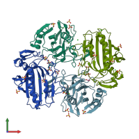
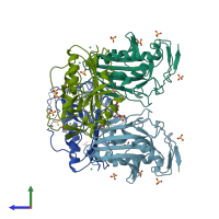
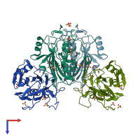
4qh0

X-ray diffraction
2Å resolution

Crystal structure of NucA from Streptococcus agalactiae with magnesium ion bound

Released:
Primary publication:
Structural characterization of the virulence factor nuclease A from Streptococcus agalactiae.
Acta Crystallogr D Biol Crystallogr 70 2937-49 (2014)
PMID: 25372684

Function and Biology Details

Biochemical function:
Biological process:
  • not assigned
Cellular component:
  • not assigned

Structure analysis Details

Assemblies composition:
monomeric (preferred)
homo dimer
PDBe Complex ID:
PDB-CPX-165097 (preferred)
Entry contents:
1 distinct polypeptide molecule
Macromolecule:
DNA-entry nuclease (Competence-specific nuclease) Chains: A, B, C, D
Molecule details ›
Chains: A, B, C, D
Length: 225 amino acids
Theoretical weight: 25.24 KDa
Source organism: Streptococcus agalactiae ILRI112
Expression system: Escherichia coli
Structure domains: Extracellular Endonuclease, subunit A

Ligands and Environments

No modified residues

Experiments and Validation Details

Entry percentile scores
X-ray source: RIGAKU MICROMAX-007 HF
Spacegroup: P63
Unit cell:
a: 123.721Å b: 123.721Å c: 157.403Å
α: 90° β: 90° γ: 120°
R-values:
R R work R free
0.158 0.155 0.19
Expression system: Escherichia coli