4r4v

X-ray diffraction
3.07Å resolution

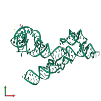
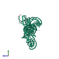
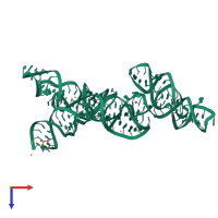
Crystal structure of the VS ribozyme - G638A mutant

Released:
Source organism: Neurospora
Primary publication:
Crystal structure of the Varkud satellite ribozyme.
Nat Chem Biol 11 840-6 (2015)
PMID: 26414446

Function and Biology Details

Biochemical function:
  • not assigned
Biological process:
  • not assigned
Cellular component:
  • not assigned

Structure analysis Details

Assembly composition:
homo dimer (preferred)
Assembly name:
PDBe Complex ID:
PDB-CPX-196864 (preferred)
Entry contents:
1 distinct RNA molecule
Macromolecule:
VS ribozyme RNA Chain: A
Molecule details ›
Chain: A
Length: 186 nucleotides
Theoretical weight: 60.17 KDa
Source organism: Neurospora
Expression system: Not provided

Ligands and Environments

2 bound ligands:
1 modified residue:

Experiments and Validation Details

Entry percentile scores
X-ray source: APS BEAMLINE 19-ID
Spacegroup: P43212
Unit cell:
a: 102.229Å b: 102.229Å c: 215.02Å
α: 90° β: 90° γ: 90°
R-values:
R R work R free
0.18 0.179 0.204
Expression system: Not provided