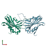
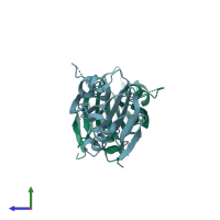
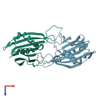
Function and Biology Details
Biochemical function:
Biological process:
Cellular component:
Sequence domains:
Structure analysis Details
Assembly composition:
monomeric (preferred)
Assembly name:
PDBe Complex ID:
PDB-CPX-165175 (preferred)
Entry contents:
1 distinct polypeptide molecule
Macromolecule:
Ligands and Environments
No bound ligands
No modified residues
Experiments and Validation Details
X-ray source:
PHOTON FACTORY BEAMLINE AR-NW12A
Spacegroup:
P1


