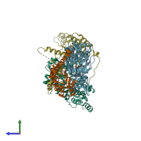
4rwt

X-ray diffraction
2.98Å resolution

Structure of actin-Lmod complex

Released:
Primary publication:
Mechanisms of leiomodin 2-mediated regulation of actin filament in muscle cells.
Proc Natl Acad Sci U S A 112 12687-92 (2015)
PMID: 26417072

Function and Biology Details

Structure analysis Details

Assembly composition:
hetero dimer (preferred)
PDBe Complex ID:
PDB-CPX-145609 (preferred)
Entry contents:
2 distinct polypeptide molecules
Macromolecules (2 distinct):
Actin-5C Chains: A, B
Molecule details ›
Chains: A, B
Length: 384 amino acids
Theoretical weight: 42.89 KDa
Source organism: Drosophila melanogaster
Expression system: Spodoptera frugiperda
UniProt:
  • Canonical: P10987 (Residues: 1-376; Coverage: 100%)
Gene names: Act5C, CG4027
Sequence domains: Actin
Structure domains:
Leiomodin-2 Chains: C, D
Molecule details ›
Chains: C, D
Length: 506 amino acids
Theoretical weight: 56.75 KDa
Source organism: Homo sapiens
Expression system: Escherichia coli
UniProt:
  • Canonical: Q6P5Q4 (Residues: 1-547; Coverage: 91%)
Gene name: LMOD2
Sequence domains: Tropomodulin
Structure domains: Ribonuclease Inhibitor

Ligands and Environments

2 bound ligands:
No modified residues

Experiments and Validation Details

Entry percentile scores
X-ray source: APS BEAMLINE 21-ID-F
Spacegroup: P1
Unit cell:
a: 65.35Å b: 65.65Å c: 81.92Å
α: 101.29° β: 90.94° γ: 107.97°
R-values:
R R work R free
0.249 0.248 0.257
Expression systems:
  • Spodoptera frugiperda
  • Escherichia coli