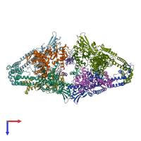
4tma

X-ray diffraction
3.3Å resolution

Crystal structure of gyrase bound to its inhibitor YacG

Released:
Model geometry
Fit model/data

Function and Biology Details

Structure analysis Details

Assembly composition:
hetero hexamer (preferred)
PDBe Complex ID:
PDB-CPX-141700 (preferred)
Entry contents:
3 distinct polypeptide molecules
Macromolecules (3 distinct):
DNA gyrase subunit A Chains: A, C, E, G
Molecule details ›
Chains: A, C, E, G
Length: 525 amino acids
Theoretical weight: 58.96 KDa
Source organism: Escherichia coli str. K-12 substr. MC4100
Expression system: Escherichia coli
Structure domains:
DNA gyrase subunit B Chains: B, D, F, H
Molecule details ›
Chains: B, D, F, H
Length: 417 amino acids
Theoretical weight: 47.34 KDa
Source organism: Escherichia coli str. K-12 substr. MC4100
Expression system: Escherichia coli BL21(DE3)
Structure domains:
DNA gyrase inhibitor YacG Chains: I, J, K, L

Ligands and Environments

1 bound ligand:
No modified residues

Experiments and Validation Details

wwPDB Validation report is not available for this entry.
X-ray source: ALS BEAMLINE 8.3.1
Spacegroup: P212121
Unit cell:
a: 107.21Å b: 114.46Å c: 462.12Å
α: 90° β: 90° γ: 90°
R-values:
R R work R free
0.245 0.243 0.283
Expression systems:
  • Escherichia coli
  • Escherichia coli BL21(DE3)