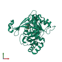
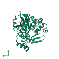
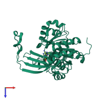
4ube

X-ray diffraction
1.93Å resolution

CRYSTAL STRUCTURE OF M TUBERCULOSIS ADENOSINE KINASE COMPLEXED WITH 2-FLURO ADENOSINE

Released:
Entry authors: Reddy MCM, Palaninathan SK, Shetty ND, Owen JL, Watson MD, Sacchettini JC

Function and Biology Details

Reaction catalysed:
ATP + adenosine = ADP + AMP
Biochemical function:
Cellular component:

Structure analysis Details

Assembly composition:
homo dimer (preferred)
Assembly name:
PDBe Complex ID:
PDB-CPX-160735 (preferred)
Entry contents:
1 distinct polypeptide molecule
Macromolecule:
Adenosine kinase Chain: A
Molecule details ›
Chain: A
Length: 334 amino acids
Theoretical weight: 35.68 KDa
Source organism: Mycobacterium tuberculosis variant bovis AF2122/97
Expression system: Escherichia coli
UniProt:
  • Canonical: P83736 (Residues: 1-324; Coverage: 100%)
Gene names: BQ2027_MB2225C, adoK, cbhK
Sequence domains: pfkB family carbohydrate kinase
Structure domains: UDP-N-acetylmuramoyl-L-alanine:D-glutamate ligase

Ligands and Environments

1 bound ligand:
No modified residues

Experiments and Validation Details

Entry percentile scores
X-ray source: RIGAKU RUH2R
Spacegroup: P3121
Unit cell:
a: 71.916Å b: 71.916Å c: 110.521Å
α: 90° β: 90° γ: 120°
R-values:
R R work R free
0.2 0.198 0.243
Expression system: Escherichia coli