


Function and Biology Details
Reaction catalysed:
(1a) [acetyl-CoA C-acyltransferase]-S-acyl-L-cyteine + acetyl-CoA = 3-oxoacyl-CoA + [acetyl-CoA C-acyltransferase]-L-cyteine
Biochemical function:
Biological process:
Cellular component:
- not assigned
Sequence domains:
Structure analysis Details
Assembly composition:
homo dimer (preferred)
Assembly name:
Steroid 3-ketoacyl-CoA thiolase (preferred)
PDBe Complex ID:
PDB-CPX-125189 (preferred)
Entry contents:
1 distinct polypeptide molecule
Macromolecule:
Steroid 3-ketoacyl-CoA thiolase
Molecule details ›
Chains: A, B
Length: 399 amino acids
Theoretical weight: 42.35 KDa
Source organism: Mycobacterium tuberculosis H37Rv
Expression system: Mycolicibacterium smegmatis MC2 155
UniProt:
Sequence domains:
Structure domains: Peroxisomal Thiolase; Chain A, domain 1
Length: 399 amino acids
Theoretical weight: 42.35 KDa
Source organism: Mycobacterium tuberculosis H37Rv
Expression system: Mycolicibacterium smegmatis MC2 155
UniProt:
- Canonical:
 I6XHI4 (Residues: 1-391; Coverage: 100%)
Sequence domains:
Structure domains: Peroxisomal Thiolase; Chain A, domain 1
Ligands and Environments


Experiments and Validation Details
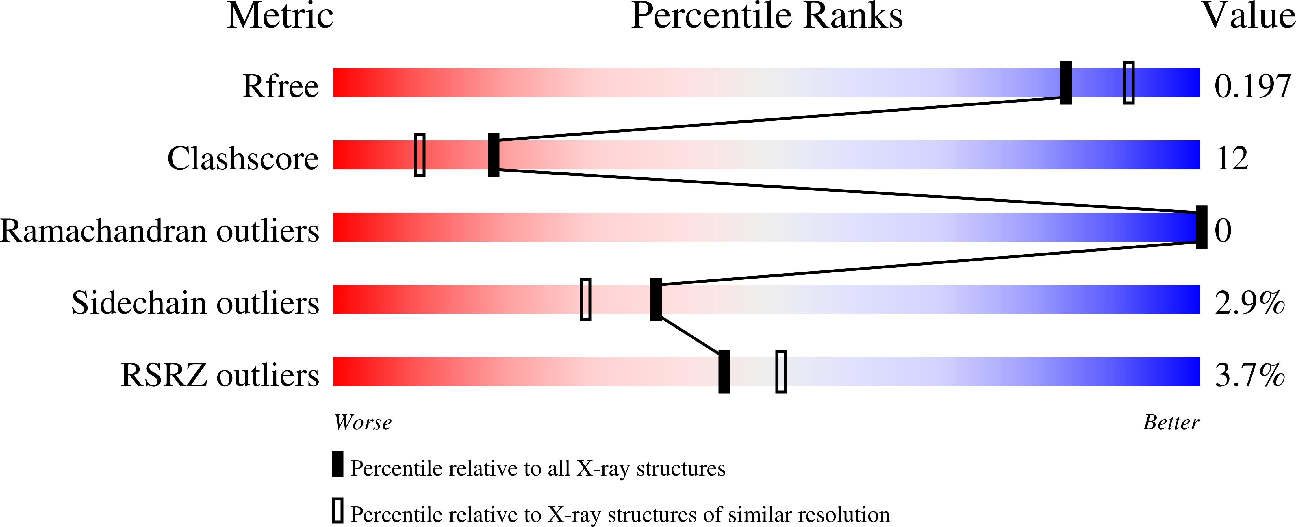
wwPDB Validation report is not available for this entry.
X-ray source:
BESSY BEAMLINE 14.1
Spacegroup:
P43212
Expression system: Mycolicibacterium smegmatis MC2 155
