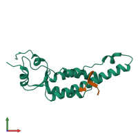
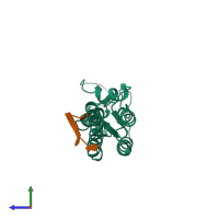
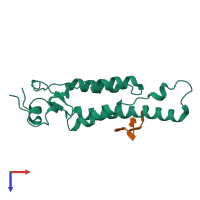
4udv

Electron Microscopy
3.35Å resolution

Cryo-EM structure of TMV at 3.35 A resolution

Released:
Source organism: Tobacco mosaic virus
Primary publication:
Seeing tobacco mosaic virus through direct electron detectors.
J Struct Biol 189 87-97 (2015)
PMID: 25528571
Related structures: EMD-2842

Function and Biology Details

Biochemical function:
Biological process:
  • not assigned
Cellular component:

Structure analysis Details

Assembly composition:
hetero 98-mer (preferred)
Assembly name:
PDBe Complex ID:
PDB-CPX-159633 (preferred)
Entry contents:
1 distinct polypeptide molecule
1 distinct RNA molecule
Macromolecules (2 distinct):
Capsid protein Chain: A
Molecule details ›
Chain: A
Length: 158 amino acids
Theoretical weight: 17.51 KDa
Source organism: Tobacco mosaic virus
UniProt:
  • Canonical: P69687 (Residues: 2-159; Coverage: 99%)
Gene name: CP
Sequence domains: Virus coat protein (TMV like)
Structure domains: Tobacco mosaic virus-like, coat protein
5'-D(*GP*AP*AP)-3' Chain: R
Molecule details ›
Chain: R
Length: 3 nucleotides
Theoretical weight: 959 Da
Source organism: Tobacco mosaic virus

Ligands and Environments

No bound ligands
No modified residues

Experiments and Validation Details

Entry percentile scores
Resolution: 3.35Å
Relevant EMDB volumes: EMD-2842