4w7c

X-ray diffraction
2.5Å resolution

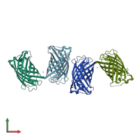
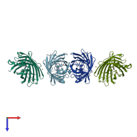
Crystal Structure of Full-Length Split GFP Mutant D21H/K26C Disulfide and Metal-Mediated Dimer, C 2 Space Group

Released:

Function and Biology Details

Biochemical function:
  • not assigned
Biological process:
Cellular component:
  • not assigned

Structure analysis Details

Assembly composition:
homo dimer (preferred)
PDBe Complex ID:
PDB-CPX-165311 (preferred)
Entry contents:
1 distinct polypeptide molecule
Macromolecule:
fluorescent protein D21H/K26C Chains: A, B, C, D
Molecule details ›
Chains: A, B, C, D
Length: 229 amino acids
Theoretical weight: 26.09 KDa
Source organism: synthetic construct
Expression system: Escherichia coli
Structure domains: Green fluorescent protein

Ligands and Environments

1 bound ligand:
1 modified residue:

Experiments and Validation Details

Entry percentile scores
X-ray source: APS BEAMLINE 24-ID-C
Spacegroup: C2
Unit cell:
a: 205.49Å b: 69.42Å c: 102.81Å
α: 90° β: 110.73° γ: 90°
R-values:
R R work R free
0.229 0.226 0.254
Expression system: Escherichia coli