

-alpha-D-glucopyranose</span>;</li> <li class='image_legend_li'>1 copy of <span class='highlight'>water</span>.</li></ul>")
-alpha-D-glucopyranose</span>;</li> <li class='image_legend_li'>1 copy of <span class='highlight'>water</span>.</li></ul>")
-alpha-D-glucopyranose</span>;</li> <li class='image_legend_li'>1 copy of <span class='highlight'>water</span>.</li></ul>")

Function and Biology Details
Reaction catalysed:
Cleavage of hydrophobic, N-terminal signal or leader sequences fromsecreted and periplasmic proteins.
Biochemical function:
Biological process:
Cellular component:
Sequence domains:
- Maltose/Cyclodextrin ABC transporter, substrate-binding protein
- Peptidase S26
- Peptidase S26A, signal peptidase I, conserved site
- Peptidase S26A, signal peptidase I, lysine active site
- LexA/Signal peptidase-like superfamily
- Peptidase S26A, signal peptidase I
- Solute-binding family 1, conserved site
- Bacterial-type extracellular solute-binding protein
Structure analysis Details
Assembly composition:
monomeric (preferred)
Assembly name:
PDBe Complex ID:
PDB-CPX-199636 (preferred)
Entry contents:
1 distinct polypeptide molecule
Macromolecules (2 distinct):
Maltose/maltodextrin-binding periplasmic protein; Signal peptidase IB
Molecule details ›
Ligands and Environments

Experiments and Validation Details
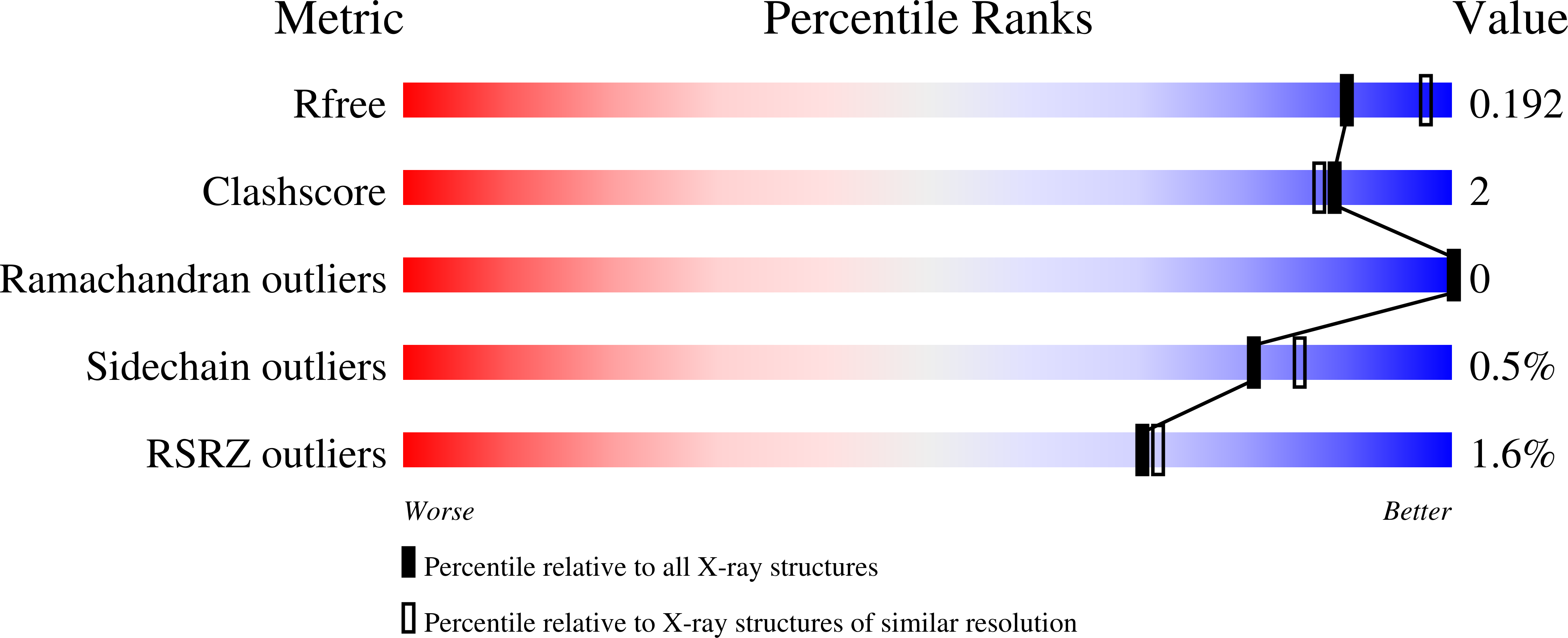
wwPDB Validation report is not available for this entry.
X-ray source:
AUSTRALIAN SYNCHROTRON BEAMLINE MX1
Spacegroup:
P21
Expression system: Escherichia coli K-12
