4y9h

X-ray diffraction
1.43Å resolution

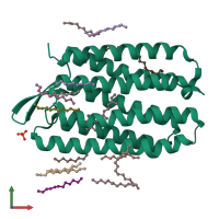
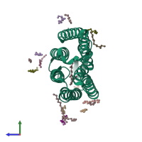
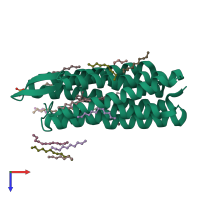
The 1.43 angstrom crystal structure of bacteriorhodopsin crystallized from bicelles

Released:
Source organism: Halobacterium salinarum R1
Entry authors: Saiki H, Sugiyama S, Kakinouchi K, Kawatake S, Hanashima S, Matsumori N, Murata M

Function and Biology Details

Structure analysis Details

Assembly composition:
monomeric (preferred)
Assembly name:
PDBe Complex ID:
PDB-CPX-109192 (preferred)
Entry contents:
1 distinct polypeptide molecule
Macromolecule:
Bacteriorhodopsin Chain: A
Molecule details ›
Chain: A
Length: 226 amino acids
Theoretical weight: 24.76 KDa
Source organism: Halobacterium salinarum R1
UniProt:
  • Canonical: B0R5N9 (Residues: 19-244; Coverage: 86%)
Gene names: OE_3106F, bop
Sequence domains: Bacteriorhodopsin-like protein
Structure domains: Rhodopsin 7-helix transmembrane proteins

Ligands and Environments

No modified residues

Experiments and Validation Details

Entry percentile scores
X-ray source: SPRING-8 BEAMLINE BL44XU
Spacegroup: C2221
Unit cell:
a: 45.215Å b: 102.794Å c: 128.337Å
α: 90° β: 90° γ: 90°
R-values:
R R work R free
0.201 0.199 0.221