


Function and Biology Details
Biochemical function:
Biological process:
- not assigned
Cellular component:
- not assigned
Sequence domains:
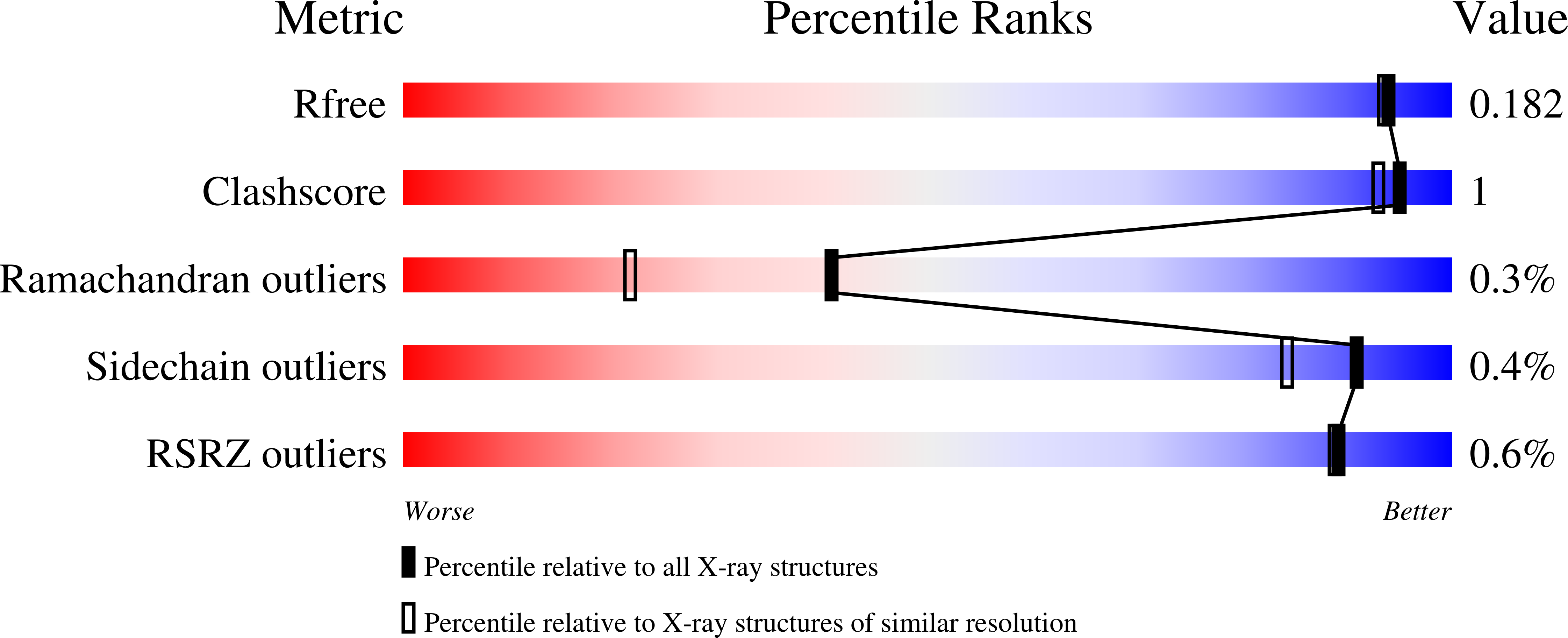
Structure analysis Details
Assemblies composition:
Assembly name:
AB hydrolase-1 domain-containing protein (preferred)
PDBe Complex ID:
PDB-CPX-108057 (preferred)
Entry contents:
1 distinct polypeptide molecule
Macromolecule:
AB hydrolase-1 domain-containing protein
Molecule details ›
Chains: A, B, C, D
Length: 304 amino acids
Theoretical weight: 33.6 KDa
Source organism: Klebsiella pneumoniae subsp. pneumoniae MGH 78578
Expression system: Escherichia coli
UniProt:
Sequence domains: alpha/beta hydrolase fold
Structure domains: alpha/beta hydrolase
Length: 304 amino acids
Theoretical weight: 33.6 KDa
Source organism: Klebsiella pneumoniae subsp. pneumoniae MGH 78578
Expression system: Escherichia coli
UniProt:
- Canonical:
 A6T9G8 (Residues: 36-338; Coverage: 100%)
Sequence domains: alpha/beta hydrolase fold
Structure domains: alpha/beta hydrolase

