


Function and Biology Details
Biochemical function:
Biological process:
Cellular component:
- not assigned
Sequence domains:
Structure analysis Details
Assemblies composition:
Assembly name:
Endoribonuclease MazF3 (preferred)
PDBe Complex ID:
PDB-CPX-161627 (preferred)
Entry contents:
1 distinct polypeptide molecule
Macromolecule:
Endoribonuclease MazF3
Molecule details ›
Chains: A, B
Length: 124 amino acids
Theoretical weight: 13.52 KDa
Source organism: Mycobacterium tuberculosis
Expression system: Escherichia coli
UniProt:
Sequence domains: PemK-like, MazF-like toxin of type II toxin-antitoxin system
Structure domains: SH3 type barrels.
Length: 124 amino acids
Theoretical weight: 13.52 KDa
Source organism: Mycobacterium tuberculosis
Expression system: Escherichia coli
UniProt:
- Canonical:
 P9WIH9 (Residues: 1-103; Coverage: 100%)
Sequence domains: PemK-like, MazF-like toxin of type II toxin-antitoxin system
Structure domains: SH3 type barrels.
Ligands and Environments
No bound ligands
No modified residues
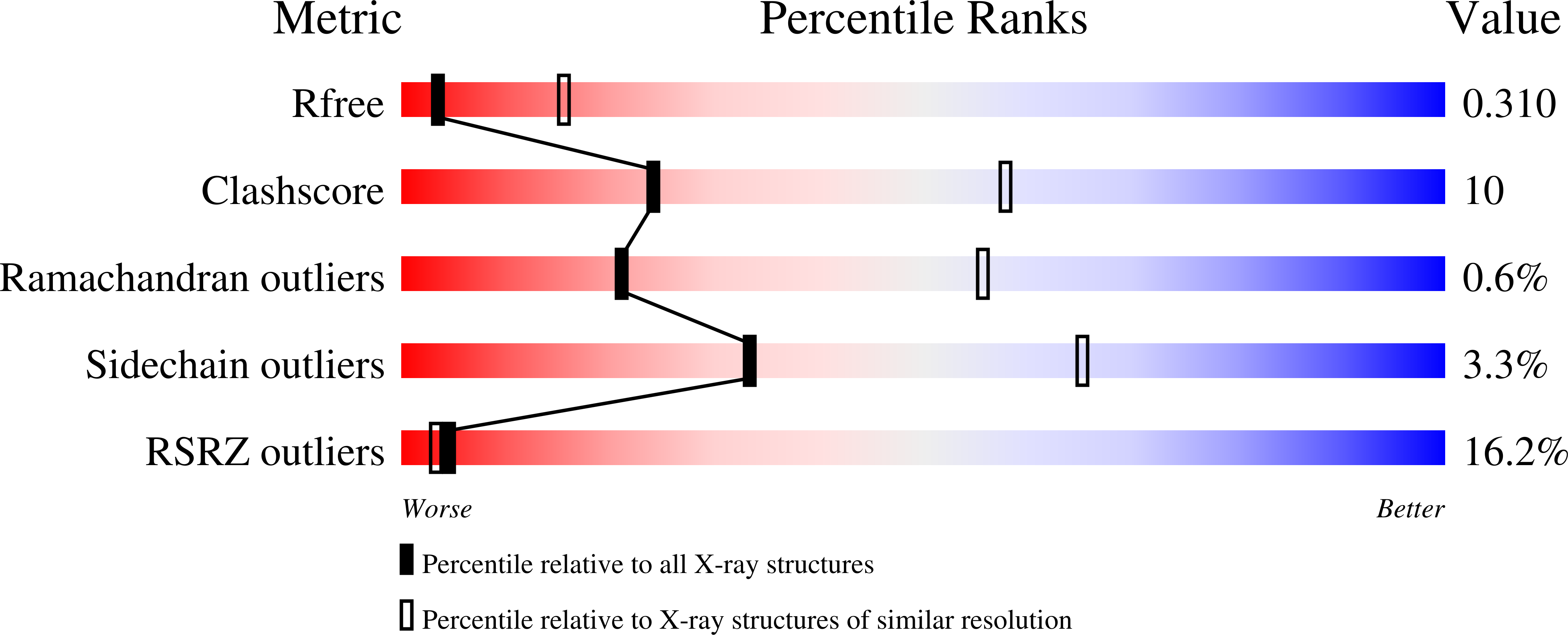
Experiments and Validation Details
wwPDB Validation report is not available for this entry.
X-ray source:
APS BEAMLINE 24-ID-C
Spacegroup:
P6422
Expression system: Escherichia coli
