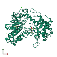
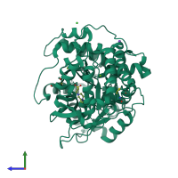
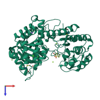
5d8g

X-ray diffraction
1.89Å resolution

A structural view on the dissociation of E. coli Tryptophanase

Released:
Source organism: Escherichia coli K-12
Primary publication:
A structural view of the dissociation of Escherichia coli tryptophanase.
Acta Crystallogr D Biol Crystallogr 71 2364-71 (2015)
PMID: 26627645

Function and Biology Details

Structure analysis Details

Assembly composition:
homo tetramer (preferred)
Assembly name:
PDBe Complex ID:
PDB-CPX-141663 (preferred)
Entry contents:
1 distinct polypeptide molecule
Macromolecule:
Tryptophanase Chain: A
Molecule details ›
Chain: A
Length: 467 amino acids
Theoretical weight: 52.5 KDa
Source organism: Escherichia coli K-12
Expression system: Escherichia coli str. 'clone D i2'
UniProt:
  • Canonical: P0A853 (Residues: 5-471; Coverage: 99%)
Gene names: JW3686, b3708, ind, tnaA
Sequence domains: Beta-eliminating lyase

Ligands and Environments

1 modified residue:

Experiments and Validation Details

Entry percentile scores
X-ray source: RIGAKU RU200
Spacegroup: F222
Unit cell:
a: 118.287Å b: 120.267Å c: 171.709Å
α: 90° β: 90° γ: 90°
R-values:
R R work R free
0.176 0.174 0.214
Expression system: Escherichia coli str. 'clone D i2'