5e22

X-ray diffraction
1.8Å resolution

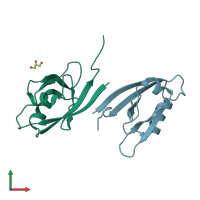
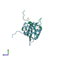
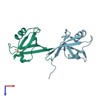
The second PDZ domain of Ligand of Numb protein X 2 in the presence of an electric field of ~1 MV/cm along the crystallographic x axis, with eightfold extrapolation of structure factor differences.

Released:
Source organism: Homo sapiens
Primary publication:
Electric-field-stimulated protein mechanics.
Nature 540 400-405 (2016)
PMID: 27926732

Function and Biology Details

Biochemical function:
  • not assigned
Biological process:
  • not assigned
Cellular component:
  • not assigned
Sequence domains:

Structure analysis Details

Assembly composition:
monomeric (preferred)
PDBe Complex ID:
PDB-CPX-185381 (preferred)
Entry contents:
1 distinct polypeptide molecule
Macromolecule:
Ligand of Numb protein X 2 Chains: A, B
Molecule details ›
Chains: A, B
Length: 95 amino acids
Theoretical weight: 10.31 KDa
Source organism: Homo sapiens
Expression system: Escherichia coli BL21(DE3)
UniProt:
  • Canonical: Q8N448 (Residues: 336-425; Coverage: 13%)
Gene names: LNX2, PDZRN1
Sequence domains: PDZ domain
Structure domains: Pdz3 Domain

Ligands and Environments

1 bound ligand:
No modified residues

Experiments and Validation Details

Entry percentile scores
X-ray source: APS BEAMLINE 14-ID-B
Spacegroup: P1
Unit cell:
a: 38.146Å b: 38.146Å c: 39.01Å
α: 113.31° β: 113.31° γ: 62.28°
R-values:
R R work R free
0.291 0.289 0.313
Expression system: Escherichia coli BL21(DE3)