


Function and Biology Details
Biochemical function:
Biological process:
Cellular component:
Structure analysis Details
Assembly composition:
monomeric (preferred)
Assembly name:
ADF-H domain-containing protein (preferred)
PDBe Complex ID:
PDB-CPX-113038 (preferred)
Entry contents:
1 distinct polypeptide molecule
Macromolecule:
ADF-H domain-containing protein
Molecule details ›
Chains: A, B
Length: 141 amino acids
Theoretical weight: 15.93 KDa
Source organism: Streptomyces griseoflavus
Expression system: Escherichia coli BL21(DE3)
UniProt:
Sequence domains: Cofilin/tropomyosin-type actin-binding protein
Structure domains: Severin
Length: 141 amino acids
Theoretical weight: 15.93 KDa
Source organism: Streptomyces griseoflavus
Expression system: Escherichia coli BL21(DE3)
UniProt:
- Canonical:
 D9XKI5 (Residues: 1-141; Coverage: 100%)
Sequence domains: Cofilin/tropomyosin-type actin-binding protein
Structure domains: Severin
Ligands and Environments
No bound ligands
No modified residues
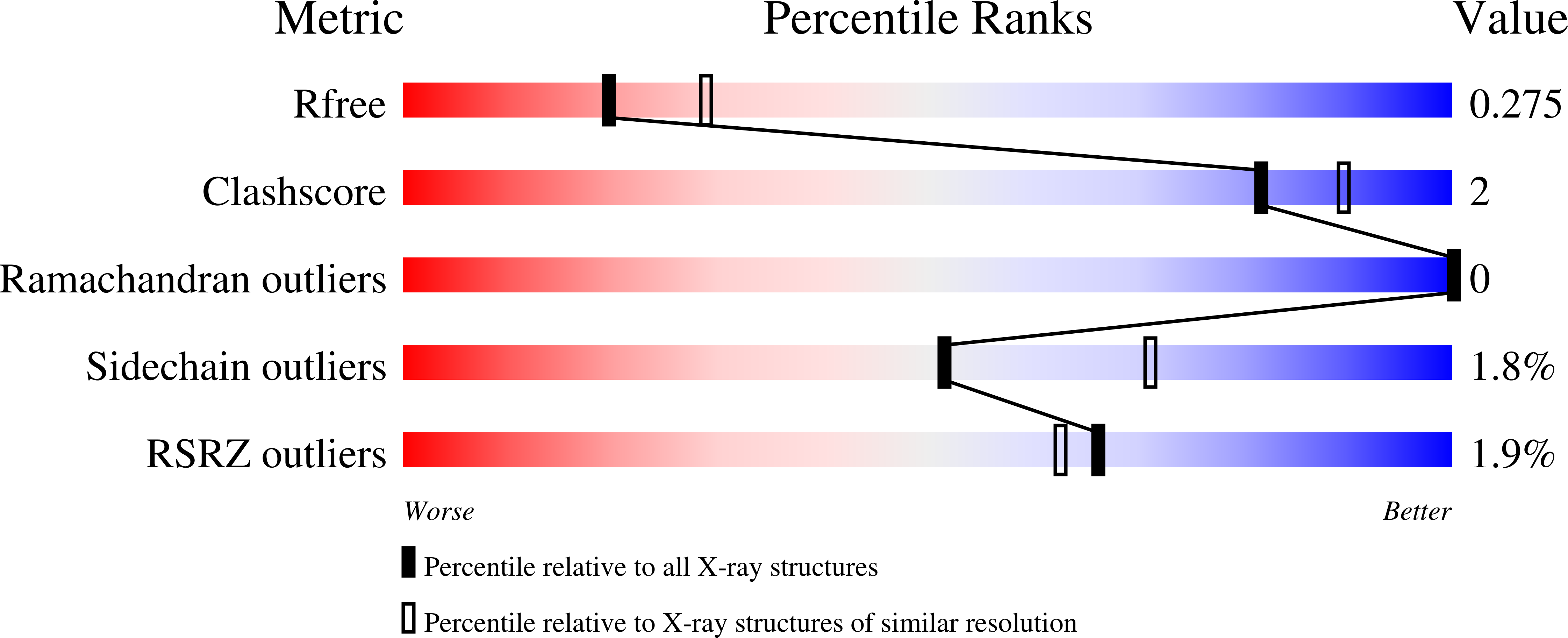
Experiments and Validation Details
wwPDB Validation report is not available for this entry.
X-ray source:
RIGAKU RU300
Spacegroup:
P212121
Expression system: Escherichia coli BL21(DE3)
