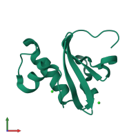
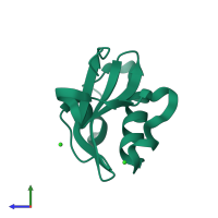
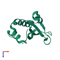
5iwh

X-ray diffraction
1.1Å resolution

Structure of P. vulgaris HigB toxin delta H92

Released:
Source organism: Proteus vulgaris
Primary publication:
Mechanism of endonuclease cleavage by the HigB toxin.
Nucleic Acids Res 44 7944-53 (2016)
PMID: 27378776

Function and Biology Details

Biochemical function:
Cellular component:
  • not assigned

Structure analysis Details

Assembly composition:
monomeric (preferred)
Assembly name:
PDBe Complex ID:
PDB-CPX-181315 (preferred)
Entry contents:
1 distinct polypeptide molecule
Macromolecule:
Endoribonuclease HigB Chain: A
Molecule details ›
Chain: A
Length: 117 amino acids
Theoretical weight: 13.58 KDa
Source organism: Proteus vulgaris
Expression system: Escherichia coli BW25113
UniProt:
  • Canonical: Q7A225 (Residues: 2-91; Coverage: 98%)
Gene name: higB
Sequence domains: RelE-like toxin of type II toxin-antitoxin system HigB
Structure domains: RelE-like

Ligands and Environments

1 bound ligand:
No modified residues

Experiments and Validation Details

Entry percentile scores
X-ray source: APS BEAMLINE 22-ID
Spacegroup: C2
Unit cell:
a: 55.776Å b: 33.166Å c: 51.492Å
α: 90° β: 115.49° γ: 90°
R-values:
R R work R free
0.14 0.138 0.169
Expression system: Escherichia coli BW25113